

The structure of a far-red fluorescent protein, AQ143, shows evidence in support of reported red-shifting chromophore interactions
- PMID: 24888769
- PMCID: PMC4116662
- DOI: 10.1002/pro.2498
The structure of a far-red fluorescent protein, AQ143, shows evidence in support of reported red-shifting chromophore interactions
Abstract
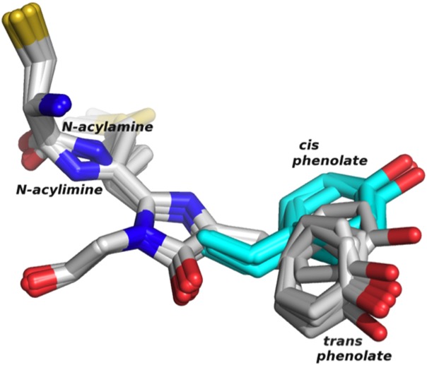
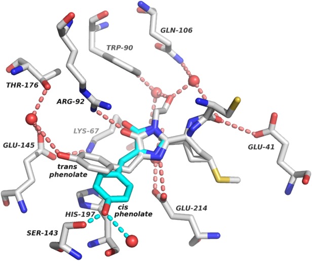
Engineering fluorescent proteins (FPs) to emit light at longer wavelengths is a significant focus in the development of the next generation of fluorescent biomarkers, as far-red light penetrates tissue with minimal absorption, allowing better imaging inside of biological hosts. Structure-guided design and directed evolution have led to the discovery of red FPs with significant bathochromic shifts to their emission. Here, we present the crystal structure of one of the most bathochromically shifted FPs reported to date, AQ143, a nine-point mutant of aeCP597, a chromoprotein from Actinia equina. The 2.19 Å resolution structure reveals several important chromophore interactions that contribute to the protein's far-red emission and shows dual occupancy of the green and red chromophores.
Keywords: chromoprotein; near-infrared, bathochromic shift; red fluorescent protein.
© 2014 The Protein Society.
Figures
References
-
- Shcherbo D, Merzlyak E, Chepurnykh T, Fradkov A, Ermakova G, Solovieva E, Lukyanov K, Bogdanova E, Zaraisky A, Lukyanov S, Chudakov D. Bright far-red fluorescent protein for whole-body imaging. Nat Methods. 2007;4:741–746. - PubMed
-
- Kredel S, Nienhaus K, Oswald F, Wolff M, Ivanchenko S, Cymer F, Jeromin A, Michel F, Spindler K-D, Heilker R, Nienhaus G, Wiedenmann J. Optimized and far-red-emitting variants of fluorescent protein eqFP611. Chem Biol. 2008;15:224–233. - PubMed
MeSH terms
Substances
Associated data
- Actions
- Actions
- Actions
- Actions
- Actions
- Actions
LinkOut - more resources
Full Text Sources
Other Literature Sources
Molecular Biology Databases
Miscellaneous
