. 2013 Oct 15;110(42):16802-7.
doi: 10.1073/pnas.1309119110. Epub 2013 Sep 30.

Structure-function analysis of ribonucleotide bypass by B family DNA replicases

Affiliations

Structure-function analysis of ribonucleotide bypass by B family DNA replicases

Anders R Clausen et al. Proc Natl Acad Sci U S A. .

Abstract

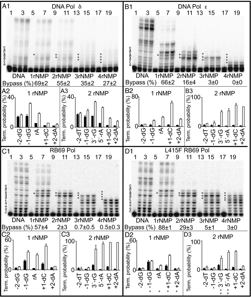
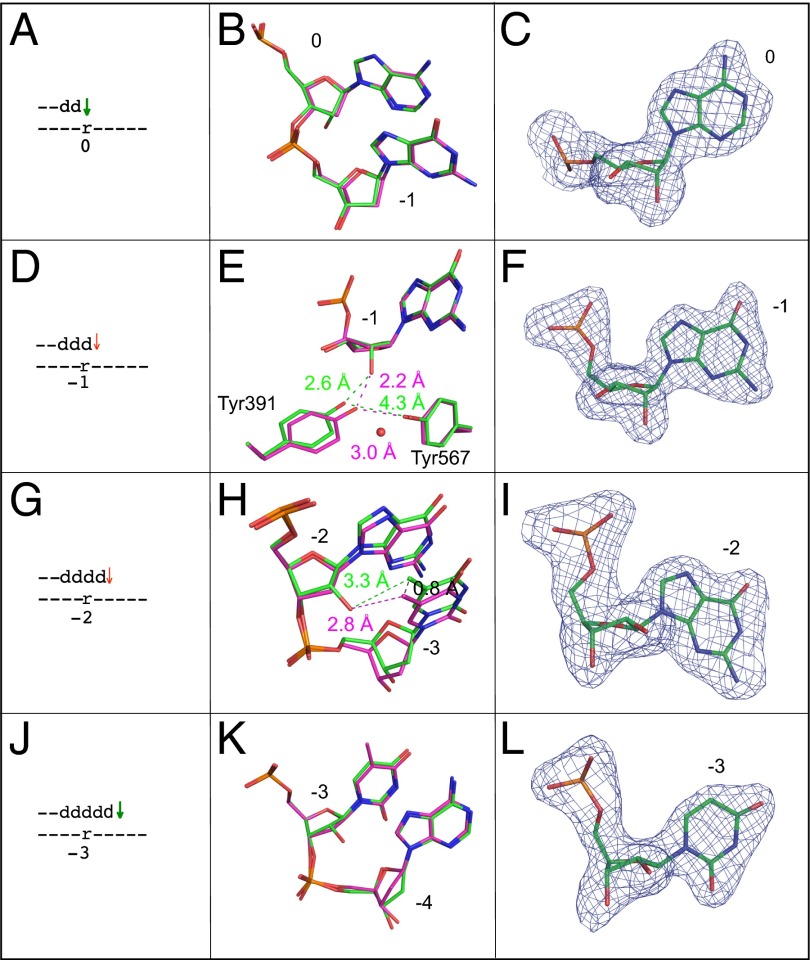
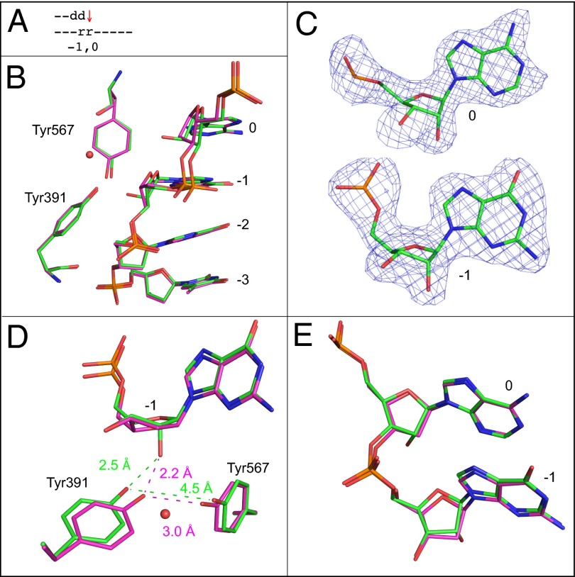
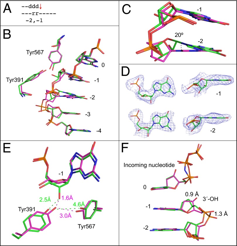
Ribonucleotides are frequently incorporated into DNA during replication, they are normally removed, and failure to remove them results in replication stress. This stress correlates with DNA polymerase (Pol) stalling during bypass of ribonucleotides in DNA templates. Here we demonstrate that stalling by yeast replicative Pols δ and ε increases as the number of consecutive template ribonucleotides increases from one to four. The homologous bacteriophage RB69 Pol also stalls during ribonucleotide bypass, with a pattern most similar to that of Pol ε. Crystal structures of an exonuclease-deficient variant of RB69 Pol corresponding to multiple steps in single ribonucleotide bypass reveal that increased stalling is associated with displacement of Tyr391 and an unpreferred C2'-endo conformation for the ribose. Even less efficient bypass of two consecutive ribonucleotides in DNA correlates with similar movements of Tyr391 and displacement of one of the ribonucleotides along with the primer-strand DNA backbone. These structure-function studies have implications for cellular signaling by ribonucleotides, and they may be relevant to replication stress in cells defective in ribonucleotide excision repair, including humans suffering from autoimmune disease associated with RNase H2 defects.

Keywords: DNA replication; replication stalling; translesion synthesis.


Conflict of interest statement

The authors declare no conflict of interest.

Figures

Fig. 1.
Fig. 1.
Bypass of ribonucleotides by Pol δ, Pol ε, and WT and L415F RB69 Pol. Bypass reactions were performed as described in Materials and Methods, in each case using a large excess of primer template over polymerase. Results are for primer extension by yeast Pol δ (A), yeast Pol ε (B), RB69 Pol (C), and L415F RB69 Pol (D). A1, B1, C1, and D1 includes PAGE phosphorimages of DNA products when copying each of five different primer templates (Table S1), from reactions incubated for 0, 4, 8, and 12 min. The efficiency of ribonucleotide bypass relative to the all-DNA control was calculated as described previously (30), and the values are shown as percentages below each set of four lanes. Termination probabilities are also shown, again calculated as described previously (30), after incorporation at each of several template positions during copying of the all-DNA template (black bars) or templates containing either one (A2, B2, C2, and D2) or two ribonucleotides (A3, B3, C3, and D3) (white bars). The asterisks in D2 correspond to the structures depicted in Fig. 2, and the double asterisks in D3 correspond to the structures depicted in Figs. 3 and 4.
Fig. 2.
Fig. 2.
Superposition of all-DNA structure (magenta) with four different single ribonucleotide structures (green). (A) Schematic depicting the ribonucleotide in the nascent base pair binding pocket (position 0). A green arrow implies that dNTP insertion is not strongly reduced, as inferred from the relatively normal termination after the preceding incorporation (Fig. 1 D1 and D2, −1-dG). (B) Overlay of the ribonucleotide-containing structure depicted in A with the all-DNA structure, showing template positions 0 and −1. (C) Simulated annealing Fo-Fc omit map contoured at 3 σ for the ribonucleotide in the nascent base pair binding pocket. (D) Stick diagram depicting the location of ribonucleotide in the primer-terminal base pair (−1 position). The red arrow implies that dNTP insertion at this position (or possibly translocation before insertion) is problematic, as indicated by the increased termination following insertion opposite rA (Fig. 1 D1 and D2, rA). (E) Overlay of the ribonucleotide containing structure depicted in D with the all-DNA structure, showing template position −1, Tyr391, and Tyr567. The red sphere indicates the position of an additional water molecule in the ribonucleotide-containing structure. (F) Simulated annealing Fo-Fc omit map contoured at 3 σ for the ribonucleotide in the −1 position. (G) Stick diagram of the position of the ribonucleotide located 2 bp upstream of the active site (−2 position). As above, the red arrow implies that dNTP insertion at this position is problematic (Fig. 1 D1 and D2, +1-dC). (H) Overlay of the ribonucleotide containing structure with the all-DNA structure at template positions −2 and −3. (I) Simulated annealing Fo-Fc omit map contoured at 3 σ for the ribonucleotide at position −2. (J) Stick diagram of the position of ribonucleotide located 3 bp upstream of the active site (−3 position). As explained above, the green arrow indicates that insertion is normal (Fig. 1 D1 and D2, +2dA). (K) Overlay of the ribonucleotide-containing structure with the all-DNA structure at template positions −3 and −4. (L) Simulated annealing Fo-Fc omit map contoured at 3 σ for the ribonucleotide at position −3.
Fig. 3.
Fig. 3.
Superposition of the all-DNA structure (magenta) with the structure containing ribonucleotides at 0 and −1 position (green). (A) Schematic indicating the positions of the two ribonucleotides. The red arrow indicates that incorporation is problematic here (Fig. 1D3, 3′-rG). (B) Superposition of template nucleotides at positions 0 to −3, Tyr391, and Tyr567. A red sphere indicates the position of an additional water molecule. (C) Simulated annealing Fo-Fc maps contoured at 3 σ for the ribonucleotides at template positions 0 and −1. (D) Overlay with the all-DNA structure, showing the ribonucleotide at the −1 position, as well as Tyr391, Tyr567, and the additional water molecule. (E) Overlay of the ribonucleotides at positions 0 and −1 with the all-DNA structure.
Fig. 4.
Fig. 4.
Superposition of the all-DNA structure (magenta) with the structure containing ribonucleotides at −1 and −2 positions (green). (A) Schematic indicating the positions of ribonucleotides at −1 and −2. The red arrow indicates that termination frequency is increased ∼10-fold compared with bypass of an all-DNA template, (Fig. 1D3, 5′-rA). (B) Superposition of template bases at 0 to −4 positions, Tyr391, and Tyr567. (C) Overlay of ribonucleotides at the −1 and −2 position with the all-DNA structure. (D) Simulated annealing Fo-Fc omit maps contoured at 3 σ are shown in blue for the two ribonucleotides at the −1 and −2 positions. (E) Overlay with the all-DNA structure showing Tyr391, Tyr567, and the nucleotide at the −1 position. (F) Overlay of primer terminus and incoming nucleotide with the all-DNA structure.

References

    1. Burgers PM. Polymerase dynamics at the eukaryotic DNA replication fork. J Biol Chem. 2009;284(7):4041–4045. - PMC - PubMed
    1. Cerritelli SM, Crouch RJ. Ribonuclease H: The enzymes in eukaryotes. FEBS J. 2009;276(6):1494–1505. - PMC - PubMed
    1. Balakrishnan L, Bambara RA. Eukaryotic lagging strand DNA replication employs a multi-pathway mechanism that protects genome integrity. J Biol Chem. 2011;286(9):6865–6870. - PMC - PubMed
    1. Nick McElhinny SA, et al. Abundant ribonucleotide incorporation into DNA by yeast replicative polymerases. Proc Natl Acad Sci USA. 2010;107(11):4949–4954. - PMC - PubMed
    1. Nick McElhinny SA, et al. Genome instability due to ribonucleotide incorporation into DNA. Nat Chem Biol. 2010;6(10):774–781. - PMC - PubMed

Publication types

MeSH terms

LinkOut - more resources