. 2013 Jul;195(14):3277-86.
doi: 10.1128/JB.00374-13. Epub 2013 May 17.

Structural basis for biofilm formation via the Vibrio cholerae matrix protein RbmA

Affiliations

Structural basis for biofilm formation via the Vibrio cholerae matrix protein RbmA

Krista M Giglio et al. J Bacteriol. 2013 Jul.

Abstract

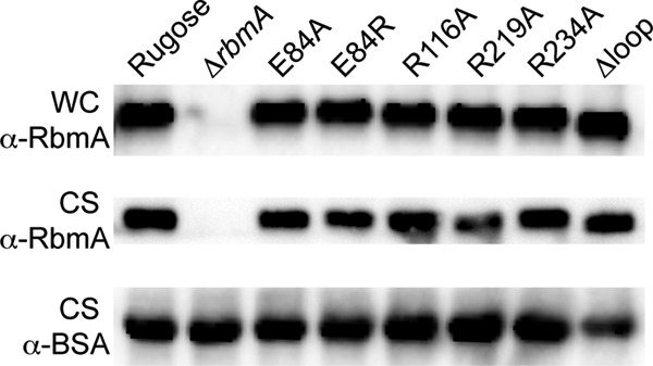
During the transition from a free-swimming, single-cell lifestyle to a sessile, multicellular state called a biofilm, bacteria produce and secrete an extracellular matrix comprised of nucleic acids, exopolysaccharides, and adhesion proteins. The Vibrio cholerae biofilm matrix contains three major protein components, RbmA, Bap1, and RbmC, which are unique to Vibrio cholerae and appear to support biofilm formation at particular steps in the process. Here, we focus on RbmA, a structural protein with an unknown fold. RbmA participates in the early cell-cell adhesion events and is found throughout the biofilm where it localizes to cell-cell contact sites. We determined crystal structures of RbmA and revealed that the protein folds into tandem fibronectin type III (FnIII) folds. The protein is dimeric in solution and in crystals, with the dimer interface displaying a surface groove that is lined with several positively charged residues. Structure-guided mutagenesis studies establish a crucial role for this surface patch for RbmA function. On the basis of the structure, we hypothesize that RbmA serves as a tether by maintaining flexible linkages between cells and the extracellular matrix.


Figures

Fig 1
Fig 1
Structure of V. cholerae RbmA. (A) Domain organization of full-length RbmA. Two molecules are shown as their domains are arranged in a crystallographic dimer. SigP, signal peptide. (B) Crystal structure. Two perpendicular views of an asymmetric unit are shown. (Top) The crystal structure revealed an RbmA dimer comprised of two tandem FnIII folds. Color coding of the individual FnIII folds is consistent with the color scheme introduced in panel A. The schematic (bottom left) depicts a half-site consisting of a FnIII fold donated by each protomer, which align in an antiparallel fashion. In the structure cartoon and the schematic, the β-strands of individual FnIII folds are labeled following a commonly used nomenclature (β-strands a to g). Unless otherwise stated, all illustrations were made with the model generated from crystal form 1 (see Table S2 in the supplemental material).
Fig 2
Fig 2
Oligomeric state of RbmA in solution. SEC-MALS was used to determine the absolute molecular mass of RbmA in solution. The light scattering signal (LS) and refractive index detector signal (dRI) are shown on the left y axis. Molecular mass determinations across the protein elution peak are shown (black dots; right y axis). The theoretical molecular weights for a dimer and monomer were calculated based on their amino acid sequence and indicated as horizontal, dashed lines. The average experimental molecular mass is 49.1 ± 1.0 kDa.
Fig 3
Fig 3
Structural neighbors of RbmA. A DALI search against the Protein Data Bank revealed structurally conserved features between RbmA (center) and, starting from the top right and going clockwise, human transglutaminase (Z score of 12.1; RMSD of 6.1 Å), a putative β-galactosidase (Z score of 10.3; RMSD of 7.0 Å), a subunit of the Mycobacterium smegmatis porin MspA (Z score of 9.0; RMSD of 2.5 Å), a Streptococcus mutans dextranase (Z score of 10.2; RMSD of 2.8 Å), and V. cholerae GbpA (Z score of 10.2; RMSD of 9.1 Å). Monomeric structures were arranged adjacent to the most homologous FnIII fold of RbmA identified in this search. The aligning domains in the individual proteins are shown in color.
Fig 4
Fig 4
Characteristics of the RbmA lobe interface. (A) Crystallographic water molecules. A large number of water molecules were resolved in the crystal structure, and many of them fill the interlobe interface within an RbmA dimer. Water molecules are shown as blue spheres. (B) Mapping of the distribution of polar and hydrophobic residues at the interlobe interface (top) and at the surface-exposed face of an individual lobe (bottom). The orientation with the molecule in panel A is shown in the circled inset.
Fig 5
Fig 5
Surface properties of RbmA. (A) Electrostatic potential. The electrostatic potential of the RbmA dimer was mapped onto its molecular surface (right models), with red representing negative potential and blue representing positive potential (−4 to + 4 kBT). Electrostatic potentials were calculated by using the program Adaptive Poisson-Boltzmann Solver (APBS). The left models are color coded as shown in Fig. 1. Two perpendicular views are shown. (B) Surface groove. A putative binding pocket at a site located at the FnIII domain dimer interface at which two protomers come together is shown as a close-up view. Residues contributing to the positive potential are shown as sticks. (C) Crystal form 1 versus crystal form 2. The alternative loop conformation of residues 91 to 108 as observed in crystal form 2 (and 3) is shown in cyan.
Fig 6
Fig 6
Low-resolution solution structure of RbmA. (A) Primary solution scattering profiles. The scattered intensity I is plotted as a function of momentum transfer s. Averaged and buffer-subtracted SAXS intensity (I) curves of RbmA solutions at 2, 5, and 10 mg/ml are shown. The radius of gyration (Rg) and the maximum dimensions of the dimeric protein (Dmax) were calculated based on the scattering data or from the crystal structure (see Results). (B) Guinier plot. The Guinier plot based on the data collected at the three different protein concentrations is shown for the low-angle region. (C) Distance distribution functions. The distance distribution functions were computed based on the scattering profile (red line shows 10-mg/ml data set) or the crystal structure of dimeric RbmA. (D) Envelope reconstructions. SAXS data were modeled ab initio using dummy residues (applying P2 symmetry during the modeling). Twenty individual models were superimposed, averaged, and filtered. The filtered envelope is shown as gray surface representation in two perpendicular views. Independently, the SAXS data were modeled using two individual lobes taken from the high-resolution RbmA structure as the input. Relative lobe orientations were restricted during the positional modeling only by distance constraints accounting for the coil-like linker segment connecting the two FnIII of an RbmA protomer. The best-fitting model was docked manually into the low-resolution envelope.
Fig 7
Fig 7
Production of RbmA in V. cholerae. Western blot analysis of RbmA production in whole-cell (WC) lysates (top blot) and secretion in culture supernatant (CS) fractions (middle blot) in rugose, ΔrbmA, and chromosomal rbmA mutants (E84A, E84R, R116A, R219A, R234A, and Δloop mutants). Equal amounts of total protein were loaded in all the blots, and BSA was used as an additional loading control for the CS fractions (bottom blot). α-RbmA, anti-RbmA antibody.
Fig 8
Fig 8
Phenotypes of chromosomal rbmA mutants. (A) Colony morphology and (B) pellicle formation of rugose, ΔrbmA, and chromosomal rbmA mutants (E84A, E84R, R116A, R219A, R234A, and Δloop mutants). The top view of pellicles formed is shown in the top row, and the side view of pellicles formed is shown in the bottom row. Bar, 0.5 mm.
Fig 9
Fig 9
Complementation of rbmA chromosomal mutant phenotypes and overexpression of rbmA point mutants. (A) Colony morphology of rugose, ΔrbmA, and chromosomal rbmA mutants (E84A, E84R, R116A, R219A, R234A, and Δloop mutants) harboring either the empty vector pMMB67EH (top row) or complementation plasmid prbmA (bottom row). Bar, 0.5 mm. (B) Colony morphologies of the rugose strain carrying the vector or the pBAD overexpression plasmids with wild-type rbmA, rbmA-R116A (the R-to-A change at position 166 encoded by rbmA), rbmA-R219A, or rbmA-R234A. Bar, 0.5 mm.

References

    1. Yildiz FH, Visick KL. 2009. Vibrio biofilms: so much the same yet so different. Trends Microbiol. 17:109–118 - PMC - PubMed
    1. Parsek MR, Singh PK. 2003. Bacterial biofilms: an emerging link to disease pathogenesis. Annu. Rev. Microbiol. 57:677–701 - PubMed
    1. Faruque SM, Albert MJ, Mekalanos JJ. 1998. Epidemiology, genetics, and ecology of toxigenic Vibrio cholerae. Microbiol. Mol. Biol. Rev. 62:1301–1314 - PMC - PubMed
    1. Alam M, Sultana M, Nair GB, Sack RB, Sack DA, Siddique AK, Ali A, Huq A, Colwell RR. 2006. Toxigenic Vibrio cholerae in the aquatic environment of Mathbaria, Bangladesh. Appl. Environ. Microbiol. 72:2849–2855 - PMC - PubMed
    1. Huq A, Sack RB, Nizam A, Longini IM, Nair GB, Ali A, Morris JG, Jr, Khan MN, Siddique AK, Yunus M, Albert MJ, Sack DA, Colwell RR. 2005. Critical factors influencing the occurrence of Vibrio cholerae in the environment of Bangladesh. Appl. Environ. Microbiol. 71:4645–4654 - PMC - PubMed

Publication types

LinkOut - more resources