. 2013 Jul 2;21(7):1182-92.
doi: 10.1016/j.str.2013.05.001. Epub 2013 Jun 20.

Crystal structures of malonyl-coenzyme A decarboxylase provide insights into its catalytic mechanism and disease-causing mutations

Affiliations

Crystal structures of malonyl-coenzyme A decarboxylase provide insights into its catalytic mechanism and disease-causing mutations

D Sean Froese et al. Structure. .

Abstract

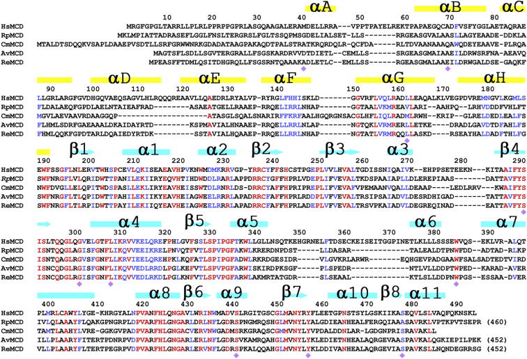
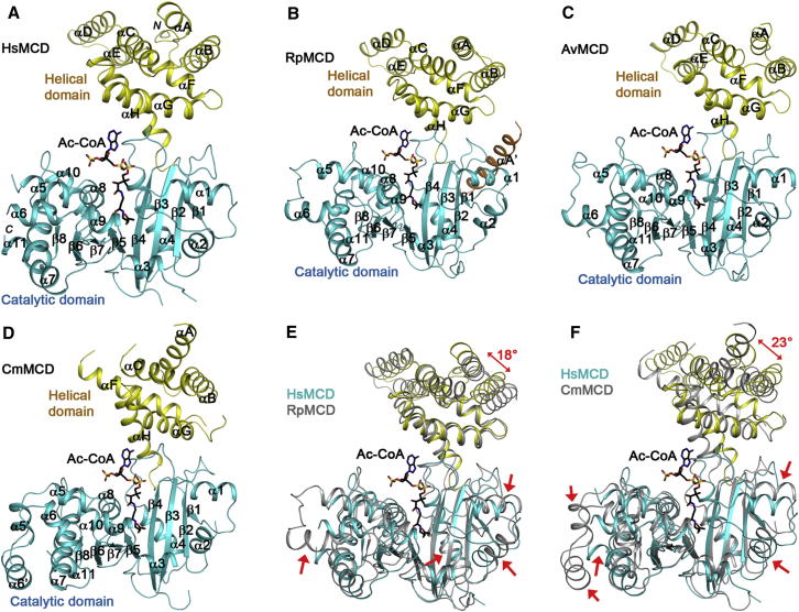
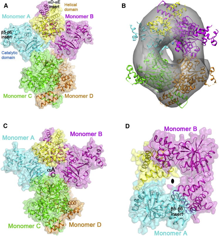
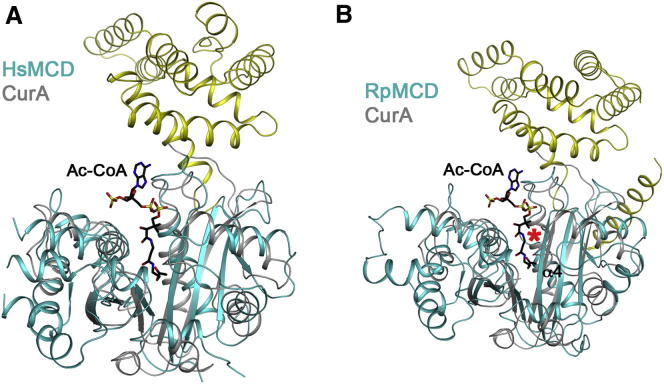
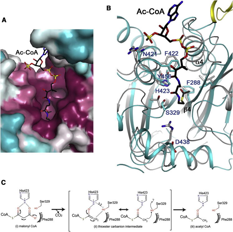
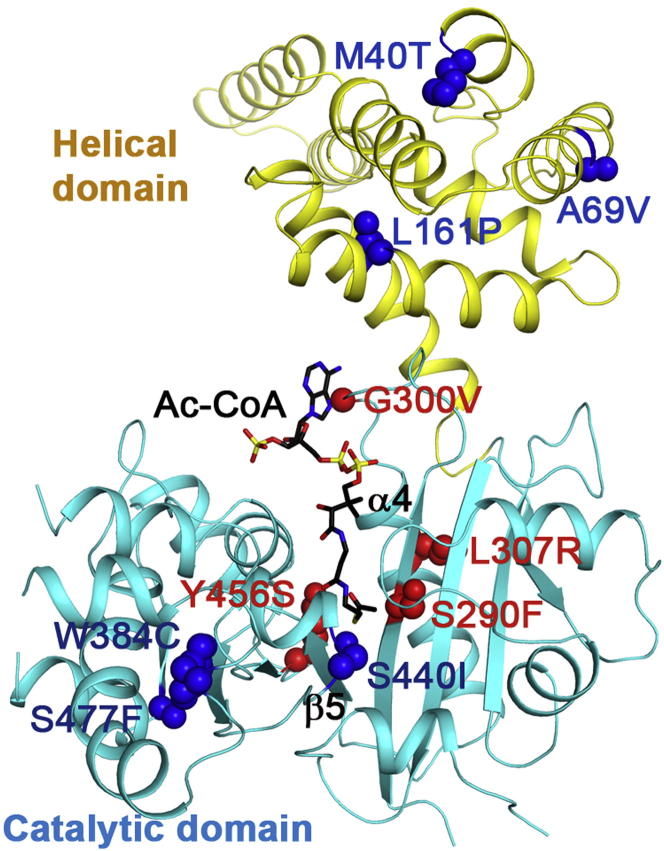
Malonyl-coenzyme A decarboxylase (MCD) is found from bacteria to humans, has important roles in regulating fatty acid metabolism and food intake, and is an attractive target for drug discovery. We report here four crystal structures of MCD from human, Rhodopseudomonas palustris, Agrobacterium vitis, and Cupriavidus metallidurans at up to 2.3 Å resolution. The MCD monomer contains an N-terminal helical domain involved in oligomerization and a C-terminal catalytic domain. The four structures exhibit substantial differences in the organization of the helical domains and, consequently, the oligomeric states and intersubunit interfaces. Unexpectedly, the MCD catalytic domain is structurally homologous to those of the GCN5-related N-acetyltransferase superfamily, especially the curacin A polyketide synthase catalytic module, with a conserved His-Ser/Thr dyad important for catalysis. Our structures, along with mutagenesis and kinetic studies, provide a molecular basis for understanding pathogenic mutations and catalysis, as well as a template for structure-based drug design.


Figures

Figure 1
Figure 1
Sequence Alignment of HsMCD, RpMCD, CmMCD, AvMCD, and ReMCD The secondary structure elements for HsMCD are indicated at the top of the alignment, colored in yellow for those in the helical domain and cyan for those in the catalytic domain. Strictly conserved residues among the five sequences are shown in red and highly conserved residues in blue. The purple diamonds indicate sites of disease-causing missense mutations in HsMCD.
Figure 2
Figure 2
Crystal Structures of MCD Monomer Schematic drawing of the structures of HsMCD (A), RpMCD (B), AvMCD (C), and CmMCD (D). The N-terminal helical domain is shown in yellow and the C-terminal catalytic domain in cyan. The bound position of acetyl-CoA in CurA (Gu et al., 2007) is shown as a stick model (in black). Overlays of the structures of HsMCD (in color) and RpMCD (in gray) (E) and HsMCD (in color) and CmMCD (in gray) (F). Regions of structural difference in the catalytic domain are highlighted with the red arrows. The difference in the orientations of the helical domains is also indicated. The structure figures were produced with PyMOL (http://www.pymol.org). See also Figure S1.
Figure 3
Figure 3
The Oligomers of MCD (A) Structure of the HsMCD tetramer. A semitransparent surface of the structure is also shown. (B) Docking of the HsMCD tetramer structure into the EM reconstruction. (C) Structure of the RpMCD tetramer. (D) Structure of the CmMCD dimer. The 2-fold axis of the dimer is indicated with the oval (black). See also Figure S2.
Figure 4
Figure 4
Structural Conservation with CurA (A) Overlay of the structures of HsMCD (in color) and CurA (in gray). Acetyl-CoA in the CurA complex is shown as a stick model (black). (B) Overlay of the structures of RpMCD (in color) and CurA (in gray). The red asterisk indicates large conformational differences in the N-terminal region of helix α4 between the two structures, which interacts with the phosphate groups of CoA in CurA.
Figure 5
Figure 5
The Active Site of MCD (A) Molecular surface of HsMCD in the active site region, colored by sequence conservation (magenta, most conserved; cyan, least conserved). The bound position of acetyl-CoA in CurA (Gu et al., 2007) is shown as a stick model (in black). (B) An overlay of HsMCD (in color) and CurA (in gray) in the active site region. Side chains in HsMCD are labeled. The catalytic residues His423 and Ser329 of HsMCD are equivalent to His389 and Thr335 of CurA. Please see Figure S3 for a stereo version of this panel. (C) Proposed catalytic mechanism for MCD (HsMCD numbering). Interatomic distance between His423 imidazole nitrogen and Ser329 hydroxyl oxygen is denoted in black line. Question mark represents possible proton transfer to reprotonate Ser329, from His423, a water molecule, or other unidentified sources. See also Figure S3.
Figure 6
Figure 6
Molecular Basis for MCD Disease-Causing Mutations The 11 missense pathogenic mutations (in red for those that could affect catalysis/substrate binding and blue for those that could affect folding/stability) are mapped onto the structure of HsMCD. See also Table S1.

References

    1. Abrahams J.P., Leslie A.G.W. Methods used in the structure determination of bovine mitochondrial F1 ATPase. Acta Crystallogr. D Biol. Crystallogr. 1996;52:30–42. - PubMed
    1. Acton T.B., Gunsalus K.C., Xiao R., Ma L.C., Aramini J., Baran M.C., Chiang Y.W., Climent T., Cooper B., Denissova N.G. Robotic cloning and protein production platform of the Northeast Structural Genomics Consortium. Methods Enzymol. 2005;394:210–243. - PubMed
    1. An J., Muoio D.M., Shiota M., Fujimoto Y., Cline G.W., Shulman G.I., Koves T.R., Stevens R., Millington D., Newgard C.B. Hepatic expression of malonyl-CoA decarboxylase reverses muscle, liver and whole-animal insulin resistance. Nat. Med. 2004;10:268–274. - PubMed
    1. Aparicio D., Pérez-Luque R., Carpena X., Díaz M., Ferrer J.C., Loewen P.C., Fita I. Structural asymmetry and disulfide bridges among subunits modulate the activity of human malonyl-CoA decarboxylase. J. Biol. Chem. 2013;288:11907–11919. - PMC - PubMed
    1. Benning M.M., Haller T., Gerlt J.A., Holden H.M. New reactions in the crotonase superfamily: structure of methylmalonyl CoA decarboxylase from Escherichia coli. Biochemistry. 2000;39:4630–4639. - PubMed

Publication types