. 2014 Jan;42(2):822-35.
doi: 10.1093/nar/gkt931. Epub 2013 Oct 22.

The double PHD finger domain of MOZ/MYST3 induces α-helical structure of the histone H3 tail to facilitate acetylation and methylation sampling and modification

Affiliations

The double PHD finger domain of MOZ/MYST3 induces α-helical structure of the histone H3 tail to facilitate acetylation and methylation sampling and modification

Ingrid Dreveny et al. Nucleic Acids Res. 2014 Jan.

Abstract

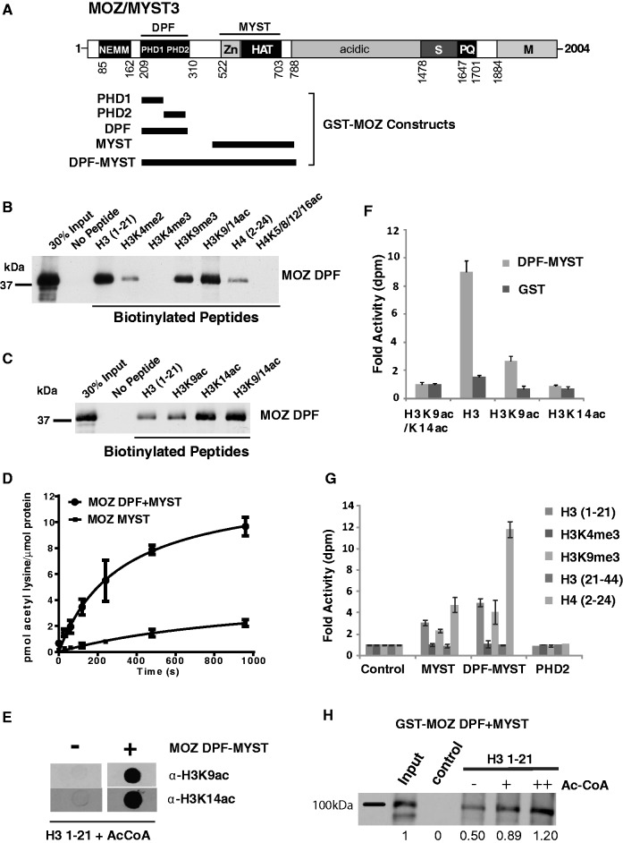
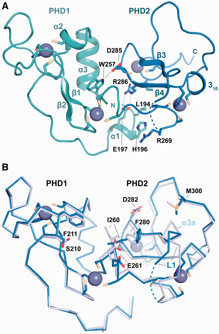
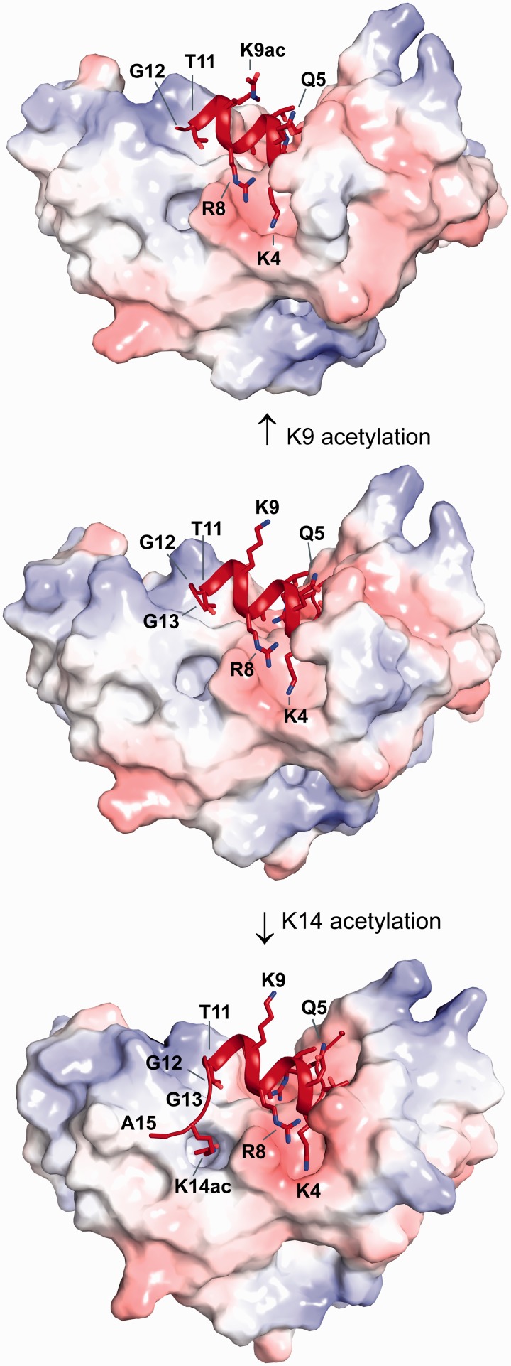
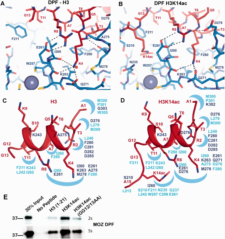
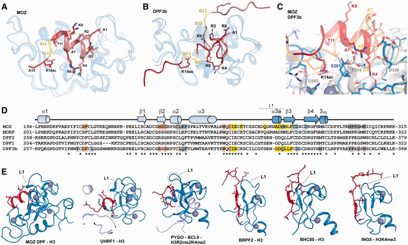
Histone tail modifications control many nuclear processes by dictating the dynamic exchange of regulatory proteins on chromatin. Here we report novel insights into histone H3 tail structure in complex with the double PHD finger (DPF) of the lysine acetyltransferase MOZ/MYST3/KAT6A. In addition to sampling H3 and H4 modification status, we show that the DPF cooperates with the MYST domain to promote H3K9 and H3K14 acetylation, although not if H3K4 is trimethylated. Four crystal structures of an extended DPF alone and in complex with unmodified or acetylated forms of the H3 tail reveal the molecular basis of crosstalk between H3K4me3 and H3K14ac. We show for the first time that MOZ DPF induces α-helical conformation of H3K4-T11, revealing a unique mode of H3 recognition. The helical structure facilitates sampling of H3K4 methylation status, and proffers H3K9 and other residues for modification. Additionally, we show that a conserved double glycine hinge flanking the H3 tail helix is required for a conformational change enabling docking of H3K14ac with the DPF. In summary, our data provide the first observations of extensive helical structure in a histone tail, revealing the inherent ability of the H3 tail to adopt alternate conformations in complex with chromatin regulators.


Figures

Figure 1.
Figure 1.
MYST DPFs function in histone acetylation and PTM recognition. (A) MOZ/MYST3 domain structure showing the NEMM (N-terminal domain of Enoki, MOZ and MORF); DPF; and MYST domains, the latter of which combines a zinc finger (Zn) and HAT domain. Regions rich in serine (S), proline and glutamine (P/Q), methionine (M) or acidic residues are also indicated. Approximate boundaries of GST-fusion proteins used for in vitro assays are also indicated. (B) MOZ DPF domains sense H3 and H4 PTMs. In vitro binding assays showing the interaction of purified GST fusion proteins with immobilized biotinylated H3 and H4 peptides, either unmodified or bearing specific modifications, as indicated. Affinity capture of recombinant GST-MOZ DPF proteins on histone peptides is detected by western blotting using α-GST antibody. (C) Effect of H3 acetylation on binding of GST MOZ DPF (D) MOZ DPF enhances histone acetylation by the MYST domain. In vitro HAT assays showing rates of acetylation of core histones by equimolar amounts of MOZ MYST domain (510–810) or the combined DPF-MYST (194–810). The data represent the mean of replicates and error bars show standard deviations. (E) MOZ DPF-MYST acetylates H3K14 and H3K9. Immunodetection of H3K9ac and H3K14ac after treatment of unmodified H3 with GST-MOZ-DPF, or GST control. (F) In vitro HAT assays showing acetylation of unmodified H3, H3K9ac and H3K14ac peptides by MOZ DPF-MYST domain, or control. (G) Acetylation of H3 and H4 peptides by MOZ PHD2, MOZ MYST or MOZ DPF-MYST proteins or control. Data columns appear in order listed in the key. (H) H3 tail acetylation enhances binding by MOZ DPF-MYST. Binding assays of GST MOZ DPF-MYST to unmodified H3 in the presence 0, 1.5 or 3 µM acetyl CoA. Proportion of affinity captured DPF-MYST proteins relative to input was quantified by densitometry.
Figure 2.
Figure 2.
Structure of the MOZ DPF domain. (A) Crystal structure of MOZ DPF spanning residues L194-G316 with the zinc atoms shown as grey spheres and the secondary structure elements indicated. The PHD1 and PHD2 subdomains are coloured in turquoise and blue, respectively. Residues engaged in interactions with the N-terminal helix α1 and PHD2 as well as a direct hydrogen bonding interaction between PHD1 and PHD2 are labelled and shown in stick representation. (B) Superposition of ribbon representations of the unbound MOZ DPF domain (blue) and the MOZ DPF domain as observed in complex with unmodified H3 (light blue); H3 is not shown for clarity. Key residues that undergo conformational changes are shown in stick representation and are labelled. Note that PHD2 is more affected by the interaction with H3 with larger changes on binding observed. Linker L1 that precedes β3 is partly disordered and the H3K4 binding pocket is partially occluded in the unbound structure.
Figure 3.
Figure 3.
Structural basis of H3, H3K9ac and H3K14ac recognition by MOZ. Surface representation of the MOZ DPF domain coloured according to electrostatic potential in complex with unmodified H3 (middle), H3K9ac (top) and H3K14ac (bottom) in red cartoon representation. Please note the exposed position of the H3K9 side chain, the preservation of the helical conformation that is independent of acetylation of either K9 or K14 and the GG hinge that mediates conformational changes to allow K14ac to bind to a pocket on the DPF domain.
Figure 4.
Figure 4.
Interactions of H3 and H3K14ac with the MOZ DPF. (A, B) Close up view of complex crystal structures in stick representation, highlighting the interactions with key residues labelled; blue denotes for MOZ DPF residues and red for H3 or H3K14ac. The DPF–H3 complex is shown in (A) and the DPF–H3K14ac complex in (B). Plausible hydrogen bonding interactions are indicated by dashed lines. Zinc atoms are shown as grey spheres. (C) H3 N-terminal tail structure as seen in the complex with MOZ DPF and (D) H3K14ac structure as seen in the complex with MOZ DPF depicted in red. Binding pockets on the DPF surface are schematically indicated as blue crescents. Corresponding interacting residues involved in hydrogen bonding interactions (dark blue) or hydrophobic contacts (light blue) are indicated. (E) Binding of GST-MOZ DPF to histone H3 tail peptides as indicated, i.e. unmodified H3, H3K14ac or H3K14ac peptide in which the GG hinge is mutated. Two exposures are shown (2 or 5 s) to highlight the differential binding.
Figure 5.
Figure 5.
Comparison of MOZ DPF–H3 with other PHD–H3 complexes. (A) Superposition of MOZ DPF (blue; semi-transparent) in complex with H3 (brown) and H3K14ac (red). The GG hinge region that mediates the insertion of K14ac into the respective binding pocket is highlighted in yellow. (B) Superposition of DPF3b in complex with unmodified H3 [PDB code: 2KWK (15)] and with H3K14ac [PDB code: 2KWJ (15)] depicted in same colour coding. In the DPF3b complex structures, the H3 conformation is extended. (C) Close up view of a superposition between MOZ DPF–H3K14ac (blue; red) and DPF3b–H3K14ac (grey; beige) (D) Sequence alignment with MOZ DPF with other DPF domain proteins. Secondary structure elements observed in our structures are shown above the alignment and MOZ residues that are engaged in H3 recognition are highlighted. Light blue denotes residues involved in H3R2 interactions, yellow for H3K4 and orange for H3K14ac interactions. Residues engaged in other interactions are depicted in grey. Colour coding is the same for DPF3b. (E) Comparison of H3 conformations in complex with assorted PHD domains. Cartoon representations of a number of DPF and PHD domains depicted in blue and H3 tail peptides depicted in red. From left to right: MOZ DPF-H3; UHRF1-H3 {PDB code 3ASK (35); PYGO-BCL9-H3R2me2K4me2 [PDB code: 2VPG; (33)]; BRPF2-H3 [PDB code: 2L43; (45)]; BHC80-H3 [PDB code 2PUY; (46)]; ING5-H3K4me3 [PDB code 3C6W; (47)}. Circled is a turn feature in the linker region L1 that may contribute to induction of helical conformation in H3 when interacting with PHD domains such as seen in MOZ, UHRF1 and potentially PYGO-BCL9.
Figure 6.
Figure 6.
(A) Histone PTM sampling by MOZ. Schematic representation showing the recruitment of MOZ proteins to chromatin through complex formation with transcription factors (TFs) or by direct interaction of MOZ with histones. Unmodified H3 and H4 tails, or modifications such as H3K9ac or H3K9me3 are permissive for MOZ recruitment. MOZ recruitment is reinforced by H3K14 acetylation. However, H3K4me3 or H4 tail acetylation constitutes non-permissive PTMs that may repulse MOZ from chromatin. (B) Histone acetylation by MOZ regulates its occupancy on chromatin. PTM sampling by the MOZ DPF facilitates histone substrate selection for acetylation. Acetylation of H3K14 stabilizes residency of MOZ on chromatin, whereas acetylation of H4 by MOZ or other regulators is non-permissive for MOZ recruitment. Histone PTMs generated by other factors such as the MLL histone methyltransferase complex that catalyses H3K4 trimethylation, also impact on MOZ residency. Thus, chromatin modifying proteins generate ‘Hire’ and ‘Fire’ PTM signatures that regulate their interactions with chromatin.

References

    1. Strahl BD, Allis CD. The language of covalent histone modifications. Nature. 2000;403:41–45. - PubMed
    1. Taverna SD, Li H, Ruthenburg AJ, Allis CD, Patel DJ. How chromatin-binding modules interpret histone modifications: lessons from professional pocket pickers. Nat. Struct. Mol. Biol. 2007;14:1025–1040. - PMC - PubMed
    1. Moriniere J, Rousseaux S, Steuerwald U, Soler-Lopez M, Curtet S, Vitte AL, Govin J, Gaucher J, Sadoul K, Hart DJ, et al. Cooperative binding of two acetylation marks on a histone tail by a single bromodomain. Nature. 2009;461:664–668. - PubMed
    1. Nady N, Lemak A, Walker JR, Avvakumov GV, Kareta MS, Achour M, Xue S, Duan S, Allali-Hassani A, Zuo X, et al. Recognition of multivalent histone states associated with heterochromatin by UHRF1 protein. J. Biol. Chem. 2011;286:24300–24311. - PMC - PubMed
    1. Ruthenburg AJ, Li H, Milne TA, Dewell S, McGinty RK, Yuen M, Ueberheide B, Dou Y, Muir TW, Patel DJ, et al. Recognition of a mononucleosomal histone modification pattern by BPTF via Multivalent Interactions. Cell. 2011;145:692–706. - PMC - PubMed

Publication types