. 2014 Mar;21(3):253-60.
doi: 10.1038/nsmb.2766. Epub 2014 Feb 2.

Sustained active site rigidity during synthesis by human DNA polymerase μ

Affiliations

Sustained active site rigidity during synthesis by human DNA polymerase μ

Andrea F Moon et al. Nat Struct Mol Biol. 2014 Mar.

Abstract

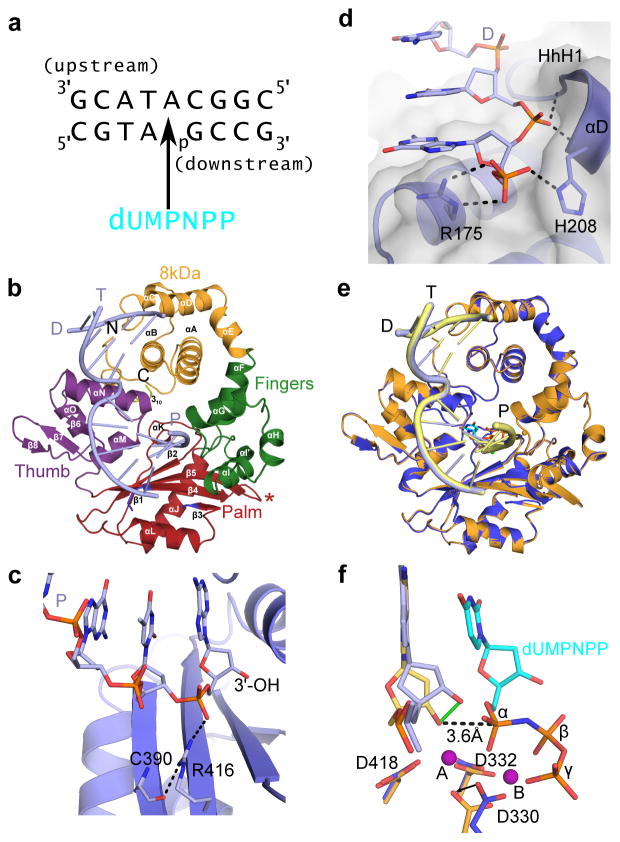
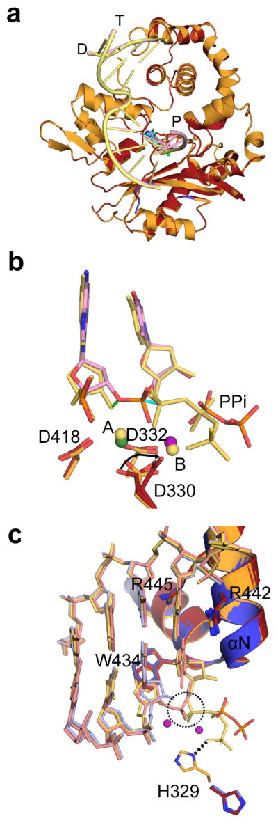
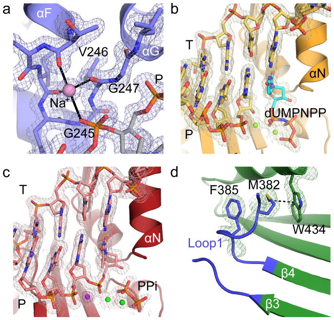
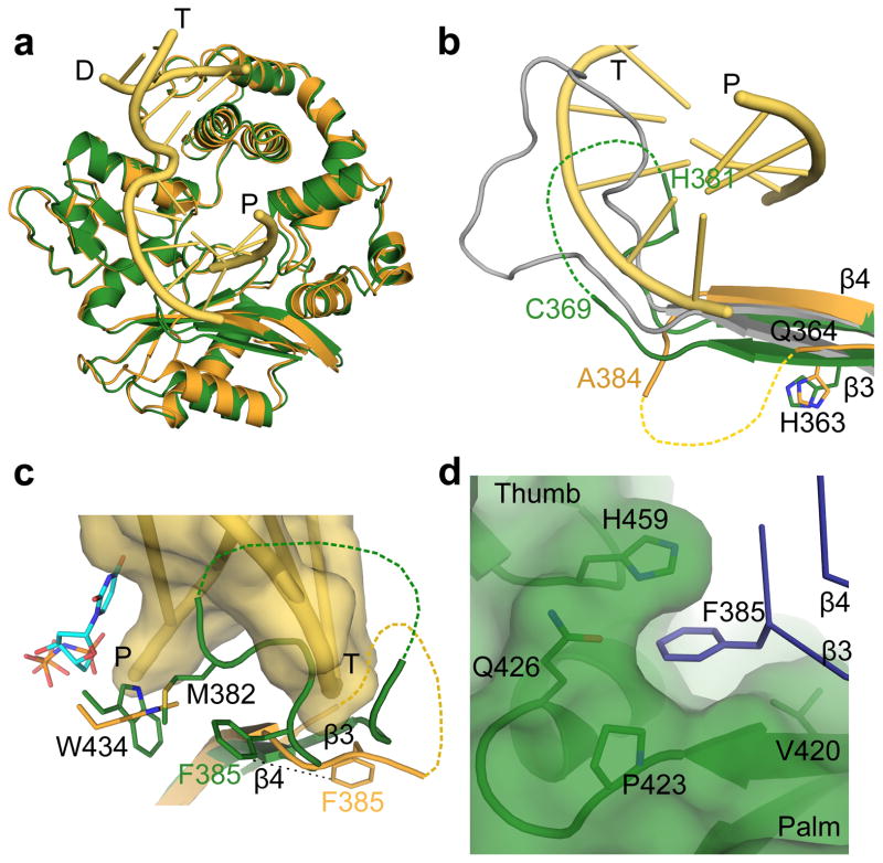
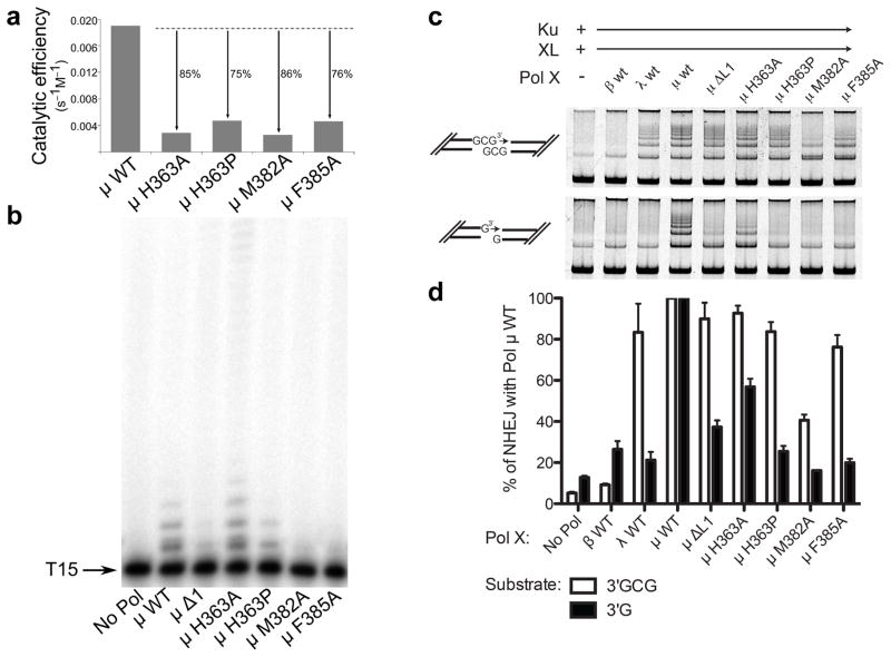
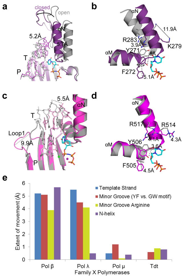
DNA polymerase μ (Pol μ) is the only template-dependent human DNA polymerase capable of repairing double-strand DNA breaks (DSBs) with unpaired 3' ends in nonhomologous end joining (NHEJ). To probe this function, we structurally characterized Pol μ's catalytic cycle for single-nucleotide incorporation. These structures indicate that, unlike other template-dependent DNA polymerases, Pol μ shows no large-scale conformational changes in protein subdomains, amino acid side chains or DNA upon dNTP binding or catalysis. Instead, the only major conformational change is seen earlier in the catalytic cycle, when the flexible loop 1 region repositions upon DNA binding. Pol μ variants with changes in loop 1 have altered catalytic properties and are partially defective in NHEJ. The results indicate that specific loop 1 residues contribute to Pol μ's unique ability to catalyze template-dependent NHEJ of DSBs with unpaired 3' ends.


Figures

Figure 1
Figure 1. Structural features of the hPol μ Δ2 binary and pre-catalytic ternary complexes
(a) Single-nucleotide gapped DNA substrate used for co-crystallization with hPol μ Δ2. (b) Ribbon diagram of the hPol μ Δ2 binary complex with substrate from (a), displaying the individual domains structures (8 kDa, fingers, palm, and thumb domains are shown in orange, green, red, and purple, respectively). Loop 1 is drawn in blue, and the truncated Loop 2 is indicated by a red asterisk. α-helices are labeled alphabetically, and β-strands numerically. The template (T), upstream primer (P), and downstream primer (D) strands are also labeled. (c) Hydrogen bonding network stabilizing the position of the primer terminus (light blue, with 3′-OH marked) in the hPol μ Δ2 binary complex (blue). (d) Binding of the 5′-phosphate on the downstream end of the gapped DNA substrate. Putative hydrogen bonding interactions between the hPol μ Δ2 8kDa subdomain (blue sticks) and the 5′-phosphorylated downstream primer (D). (e) Structural superposition of the hPol μ Δ2 binary (blue, DNA in light blue) and pre-catalytic ternary complexes (orange, DNA in yellow). (f) Superposition of the catalytic centers of the binary and ternary complexes (colored as in e). Movement of the primer terminal 3′-OH upon binding of the incoming nucleotide is shown as a solid green line. The distance between the primer terminal 3′-OH and the α-phosphate is shown as a dashed line. Mg2+ ions are shown as purple spheres. All structural figures were generated using PyMOL (Schrödinger, http://www.pymol.org).
Figure 2
Figure 2. Structural characterization of the nucleotide incorporation by hPol μ Δ2
(a) Structural superposition of the hPol μ Δ2 pre-catalytic ternary (orange, DNA in yellow) and nicked (red, DNA in pink). (b) Superposition of the primer terminal and incoming nucleotides from the pre-catalytic hPol μ Δ2 ternary complex (yellow) with the newly incorporated base from the post-catalytic complex (pink). Movement of the primer terminal 3′-OH and the α-phosphates are indicated by green and cyan solid lines, respectively. Divalent metals from the ternary complex are shown as yellow spheres, while those from the post-catalytic complex are shown in green (Mn2+) and purple (Mg2+). (c) Superposition of the active centers of the binary (blue, DNA in light blue), pre-catalytic ternary (orange, DNA in yellow) and post-catalytic nicked (red, DNA in pink) complexes of hPol μ Δ2. Incorporation of the incoming nucleotide leads to inversion of the α-phosphate (dashed circle) and release of inorganic pyrophosphate.
Figure 3
Figure 3. Electron density for hPol μ Δ2 structures
2Fo-Fc electron density maps, contoured at 1σ, for (a) sodium ion coordinated by HhH2 in the hPol μ Δ2 binary complex, active site regions of the (b) pre-catalytic ternary complex, and (c) the post-catalytic complex. The Mg2+ ions are drawn as green spheres, the incoming nucleotide in the ternary complex is shown in cyan, and the pyrophosphate released from phosphodiester bond formation in orange. The anomalous difference map (violet) for the Mn2+ (dark purple) in the active site of the post-catalytic complex is contoured to 5σ. (d) Position and interactions involving Loop1 in the hPol μ Δ2 apoprotein.
Figure 4
Figure 4. Structural characteristics of the hPol μ apoprotein
(a) Structural superposition of the hPol μ Δ2 apoprotein (green) with the pre-catalytic ternary (orange, DNA in yellow) complex. (b) Comparison of Loop1 conformations in hPol μ Δ2 apoprotein (green) and pre-catalytic ternary (orange, DNA in yellow), compared to the structurally homologous region in murine TdT (PDB ID code 1JMS, gray) structures. Ordered residues at either end of Loop 1 are labeled. Hypothetical positions of disordered sections of the loop are shown as dashed lines. (c) Position of Loop1 in the hPol μ apoprotein (green), in relation to the bound single-nucleotide gapped DNA substrate (yellow) and incoming nucleotide (cyan) from the hPol μ Δ2 ternary complex (orange). (d) Location and composition of the binding pocket containing Phe385 in the hPol μ Δ2 apoprotein structure.
Figure 5
Figure 5. Biochemical characterization of wildtype and Loop1 mutants of hPol μ
(a) Comparison of the catalytic efficiencies of hPol μ Loop1 mutants (3 nM), as determined by steady-state kinetics for template-dependent single-nucleotide incorporation. (b) Template-independent synthesis of wildtype and hPol μ Loop1 mutants (100 nM) on a single-stranded oligo dT15 DNA substrate. (c) Activity of hPol μ Loop1 mutants during in vitro NHEJ of DNA ends with partly complementary overhangs (top panel), or noncomplementary overhangs (bottom panel). Ku, XRCC4-ligase IV complex, and polymerase mutants (0.5nM) were added to linear substrates with end structures varied as noted in cartoons beside each panel. (d) Comparison of NHEJ end joining activity for hPol μ mutants, for either complementary (white bars) or noncomplementary (black bars) substrates, relative to joining observed in the presence of wildtype hPol μ. Error bars represent the standard error of the mean, for reactions performed in triplicate. Images of uncropped gels used in this study are found in Supplementary Fig. 5.
Figure 6
Figure 6. Comparison of structural rigidity across the Family X polymerases
(a) Superposition of binary (PDB ID code 3IBS, gray; DNA in light gray) and pre-catalytic ternary (PDB ID code 2FMS, purple; DNA in lavender) complexes of human Pol β, focusing on movement of the thumb subdomain and DNA strands upon incoming nucleotide (cyan) binding. (b) Side chain rearrangements occurring in the Pol β active site as a consequence of nucleotide binding. Dashed lines illustrate ‘open’ to ‘closed’ transitions. (c) Superposition of binary (PDB ID code 1XSL, gray; DNA in light gray) and pre-catalytic ternary (PDB ID code 2PFO, magenta; DNA in pink) complexes of human Pol λ. Movements of the DNA template strand (T) and Loop1 are emphasized. (d) Active site sidechain rearrangements on Pol λ α-helices M and N in preparation for catalysis. Perspective matches that for Pol β in panel (d). (e) Comparison of structural motion in Family X polymerases during active site assembly. inary and pre-catalytic ternary structures of Pol β (binary PDB ID code 3ISB, ternary PDB ID code 2FMS), Pol λ (binary PDB ID code 1XSL, ternary PDB ID code 2PFO), Pol μ, and TdT (PDB ID codes 4I2A and 4I27) were superimposed, using the structurally conserved Cα atoms of the palm subdomain (see Online Methods for details of structural analysis). The extent of protein subdomain motion (purple bar), minor groove residues (red bar), the N-helix (orange bar), and DNA template strand (blue bar) movements were calculated in Å, using the measurement wizard in PyMOL (Schrödinger, http://www.pymol.org).

References

    1. Moon AF, et al. The X family portrait: structural insights into biological functions of X family polymerases. DNA Repair (Amst) 2007;6:1709–25. - PMC - PubMed
    1. Nick McElhinny SA, Ramsden DA. Sibling rivalry: competition between Pol X family members in V(D)J recombination and general double strand break repair. Immunol Rev. 2004;200:156–64. - PubMed
    1. DeRose EF, et al. Solution structure of polymerase mu’s BRCT Domain reveals an element essential for its role in nonhomologous end joining. Biochemistry. 2007;46:12100–10. - PMC - PubMed
    1. Mueller GA, et al. A comparison of BRCT domains involved in nonhomologous end-joining: introducing the solution structure of the BRCT domain of polymerase lambda. DNA Repair (Amst) 2008;7:1340–51. - PMC - PubMed
    1. Bork P, et al. A superfamily of conserved domains in DNA damage-responsive cell cycle checkpoint proteins. FASEB J. 1997;11:68–76. - PubMed

Publication types