. 2014 May;42(9):6052-63.
doi: 10.1093/nar/gku234. Epub 2014 Apr 1.

Crystal structure of Saccharomyces cerevisiae mitochondrial GatFAB reveals a novel subunit assembly in tRNA-dependent amidotransferases

Affiliations

Crystal structure of Saccharomyces cerevisiae mitochondrial GatFAB reveals a novel subunit assembly in tRNA-dependent amidotransferases

Yuhei Araiso et al. Nucleic Acids Res. 2014 May.

Abstract

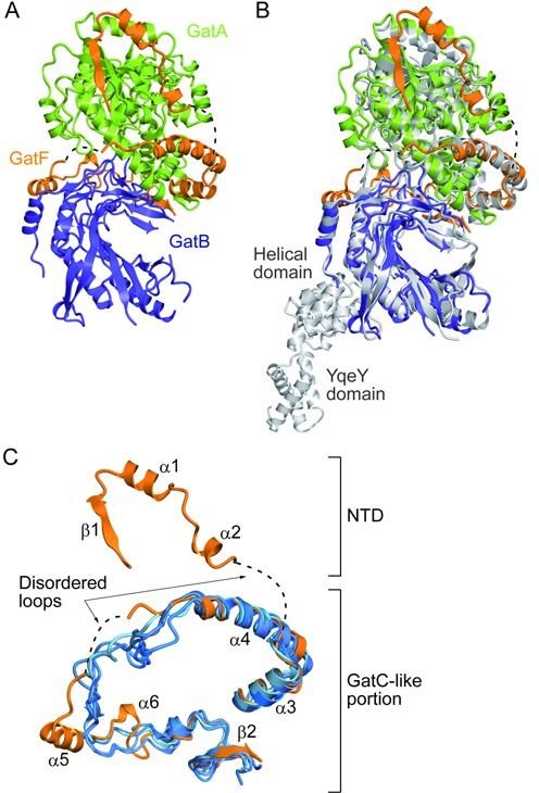
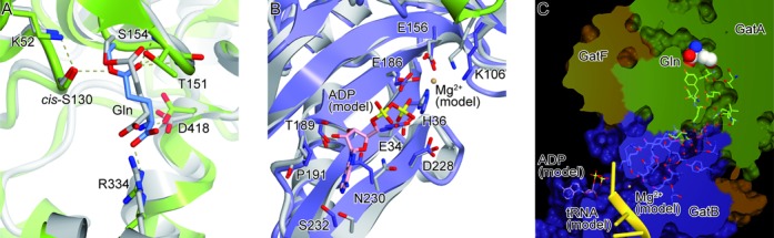
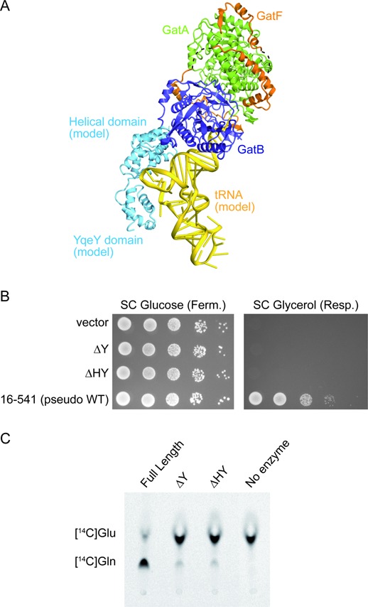
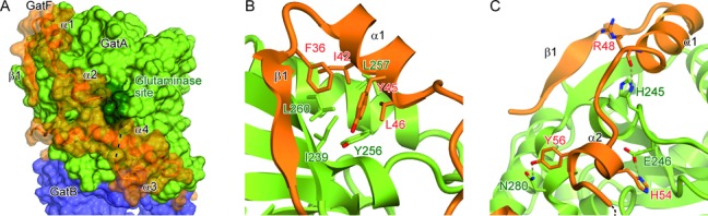
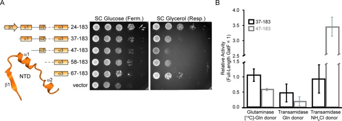
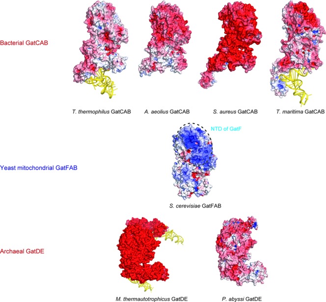
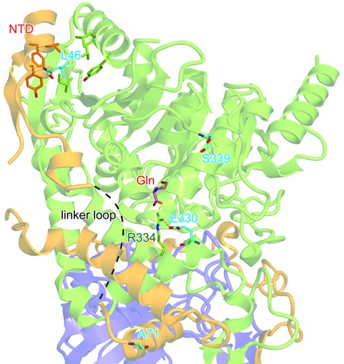
Yeast mitochondrial Gln-mtRNAGln is synthesized by the transamidation of mischarged Glu-mtRNAGln by a non-canonical heterotrimeric tRNA-dependent amidotransferase (AdT). The GatA and GatB subunits of the yeast AdT (GatFAB) are well conserved among bacteria and eukaryota, but the GatF subunit is a fungi-specific ortholog of the GatC subunit found in all other known heterotrimeric AdTs (GatCAB). Here we report the crystal structure of yeast mitochondrial GatFAB at 2.0 Å resolution. The C-terminal region of GatF encircles the GatA-GatB interface in the same manner as GatC, but the N-terminal extension domain (NTD) of GatF forms several additional hydrophobic and hydrophilic interactions with GatA. NTD-deletion mutants displayed growth defects, but retained the ability to respire. Truncation of the NTD in purified mutants reduced glutaminase and transamidase activities when glutamine was used as the ammonia donor, but increased transamidase activity relative to the full-length enzyme when the donor was ammonium chloride. Our structure-based functional analyses suggest the NTD is a trans-acting scaffolding peptide for the GatA glutaminase active site. The positive surface charge and novel fold of the GatF-GatA interface, shown in this first crystal structure of an organellar AdT, stand in contrast with the more conventional, negatively charged bacterial AdTs described previously.


Figures

Figure 1.
Figure 1.
Structural comparison of GatFAB with GatCAB. (A) Ribbon representation of the crystal structure of S. cerevisiae GatFAB, consisting of the GatA (green), GatB (navy blue) and GatF (orange) subunits. Dashed lines indicate the disordered loops of GatF (residues 59–66 and 114–128). (B) The superposition of the crystal structure of S. cerevisiae GatFAB onto that of S. aureus GatCAB. GatFAB is shown in the same color code as in (A). S. aureus GatCAB is colored gray (PDB ID: 3IP4). (C) Structural comparison of S. cerevisiae GatF with the bacterial GatC. GatF is colored orange. The T. thermophilus, T. maritima, S. aureus and A. aeolicus GatCAB structures (PDB IDs: 3KFU, 3AL0, 3IP4 and 3H0R) are colored blue and superposed onto that of S. cerevisiae GatFAB.
Figure 2.
Figure 2.
Active sites of GatFAB. GatFAB is colored as in Figure 1. (A) The glutaminase site of GatA in the Gln-bound form. The bound Gln molecule is shown as blue sticks. The crystal structure of A. aeolius GatA is colored gray and superposed onto that of S. cerevisiae GatA. (B) The amidotransferase site of GatB in the apo form. The crystal structure of S. aureus GatB is colored gray and superposed onto that of S. cerevisiae GatB. The modeled ADP molecule and Mg2+ ion are shown as pink sticks and as a sphere, respectively. (C) Surface model representation of a hydrophilic NH3 tunnel. The tunnel is filled with water molecules (red spheres), which interact with conserved residues of GatA and GatB. The modeled ADP and Mg2+ ion are shown as in (B). The modeled T. thermophilus tRNAAsn is colored yellow.
Figure 3.
Figure 3.
Functional analyses of YqeY and the helical domain of GatB. (A) Model of the GatFAB-tRNA complex. tRNAAsn from T. thermophilus (PDB ID: 3KFU) and the helical and YqeY domains of GatB from T. maritima (PDB ID: 3AL0) are docked onto the GatFABΔHY structure. The modeled tRNAAsn and the C-terminal domains of GatB are colored yellow and cyan, respectively. GatFAB is colored as in Figure 1. (B) Growth phenotypes of GatB mutants on fermentative (SC Glucose) or respiratory (SC Glycerol) media. (C) Bioimager scan of the TLC plate for a transamidase assay, which shows the absence of conversion of H. pylori tRNAGln-bound [14C]Glu into [14C]Gln, catalyzed by the GatFAB mutants with the C-terminal deletions of GatB (ΔY and ΔHY).
Figure 4.
Figure 4.
Interface between GatA and the NTD of GatF. GatFAB is colored as in Figure 1. (A) Surface representation of GatFAB. GatF is also shown in a ribbon representation. The glutaminase site is colored dark green. (B and C) Close-up views of the GatF–GatA interface.
Figure 5.
Figure 5.
Functional analyses of the GatF NTD. (A) Secondary structures of GatF mutants are shown (left). Growth phenotypes of GatF mutants on fermentative (SC Glucose) or respiratory (SC Glycerol) media (right). (B) Quantification of the activities of the GatFABF37–183 and GatFABF47–183 mutants relative to the full-length GatFAB. Glutaminase activity as well as transamidase activities with glutamine or NH4Cl as the amide donor are shown for GatF mutants relative to the activity of the full-length GatFAB. Results are the average of three experiments, with error bars representing standard deviation (See Supplementary Figure S5).
Figure 6.
Figure 6.
Electrostatic surface potential models of tRNA-dependent amidotransferases. Bacterial GatCABs (PDB IDs: 3KFU for T. thermophilus GatCAB-tRNAAsn complex, 3H0R for A. aeolius GatCAB, 3IP4 for S. aureus GatCAB and 3AL0 for T. maritima GatCAB-tRNAGln complex) are shown on the upper lane. S. cerevisiae GatFAB (this study) is shown on the center of the figure. The NTD of GatF is indicated by the dashed circle. Archaeal GatDEs (PDB IDs: 2D6F for M. thermautotrophicus GatDE-tRNAGln complex and 1ZQ1 for P. abyssi GatDE) are shown on the lower lane. Electrostatic surface potential is calculated by the program APBS (36).
Figure 7.
Figure 7.
Structural mapping of respiration-defective mutations. GatFAB is shown as in Figure 1. The residues associated with the respiration-defective phenotypes are shown as cyan sticks. The bound Gln molecule is shown as pink sticks.

References

    1. Bonnefond L., Fender A., Rudinger-Thirion J., Giegé R., Florentz C., Sissler M. Toward the full set of human mitochondrial aminoacyl-tRNA synthetases: characterization of AspRS and TyrRS. Biochemistry. 2005;44:4805–4816. - PubMed
    1. Frechin M., Duchêne A.M., Becker H.D. Translating organellar glutamine codons: a case by case scenario? RNA Biol. 2009;6:31–34. - PubMed
    1. Sagan L. On the origin of mitosing cells. J. Theor. Biol. 1967;14:255–274. - PubMed
    1. Curnow A.W., Tumbula D.L., Pelaschier J.T., Min B., Söll D. Glutamyl-tRNA(Gln) amidotransferase in Deinococcus radiodurans may be confined to asparagine biosynthesis. Proc. Natl. Acad. Sci. U.S.A. 1998;95:12838–12843. - PMC - PubMed
    1. Tumbula D.L., Becker H.D., Chang W.Z., Söll D. Domain-specific recruitment of amide amino acids for protein synthesis. Nature. 2000;407:106–110. - PubMed

Publication types

MeSH terms

Substances