. 2014:5:3004.
doi: 10.1038/ncomms4004.

Structural analysis of human 2'-O-ribose methyltransferases involved in mRNA cap structure formation

Affiliations
Free PMC article

Structural analysis of human 2'-O-ribose methyltransferases involved in mRNA cap structure formation

Miroslaw Smietanski et al. Nat Commun. 2014.
Free PMC article

Abstract

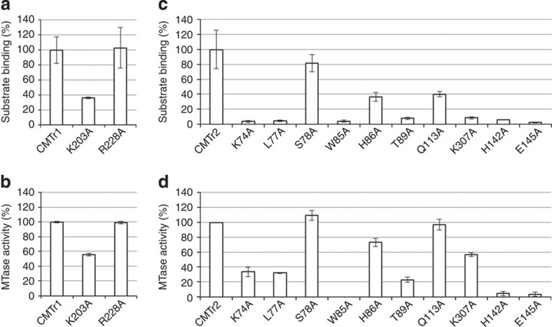
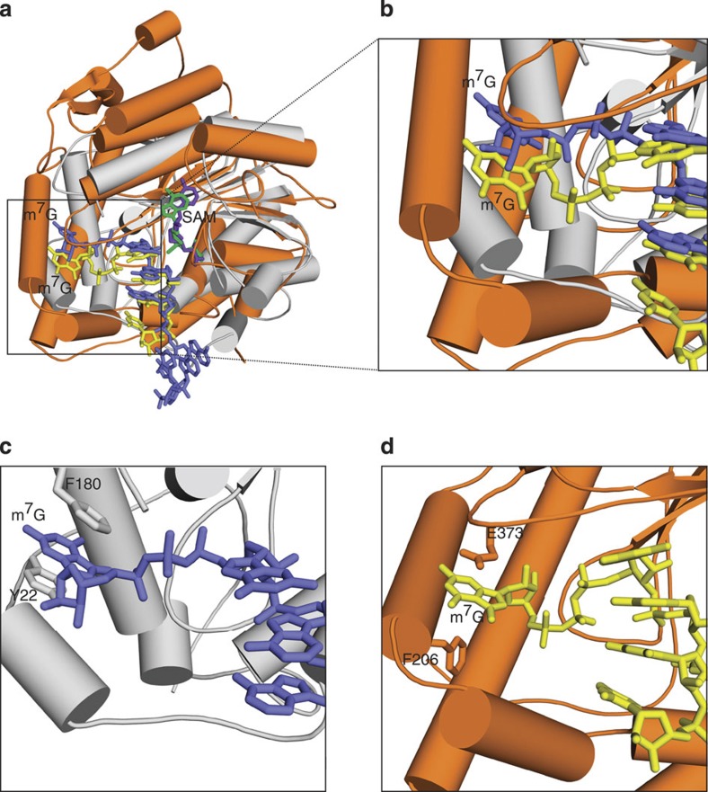
The 5' cap of human messenger RNA contains 2'-O-methylation of the first and often second transcribed nucleotide that is important for its processing, translation and stability. Human enzymes that methylate these nucleotides, termed CMTr1 and CMTr2, respectively, have recently been identified. However, the structures of these enzymes and their mechanisms of action remain unknown. In the present study, we solve the crystal structures of the active CMTr1 catalytic domain in complex with a methyl group donor and a capped oligoribonucleotide, thereby revealing the mechanism of specific recognition of capped RNA. This mechanism differs significantly from viral enzymes, thus providing a framework for their specific targeting. Based on the crystal structure of CMTr1, a comparative model of the CMTr2 catalytic domain is generated. This model, together with mutational analysis, leads to the identification of residues involved in RNA and methyl group donor binding.


Figures

Figure 1
Figure 1. Crystal structure of the catalytic MTase domain of CMTr1.
(a) Domain composition of full-length CMTr1. The dashed lines indicate the region of the protein (CMTr1126–550) present in the crystal structure. The domain boundaries are indicated with residue numbers. (b) Crystal structure of CMTr1126–550 in complex with capped oligoribonucleotide (m7GpppGAUC; coloured yellow) and SAM (green). Helices are shown in orange, β-strands are shown in blue and loops are shown in white.
Figure 2
Figure 2. Homology model of CMTr21–423.
(a) Domain composition of CMTr2. The dashed lines indicate a part of the protein that was modelled based on the CMTr1126–550 crystal structure. The domain boundaries are indicated with residue numbers. (b) Homology model of the catalytic domain of CMTr2 in complex with capped oligoribonucleotide (m7GpppGGAA) and SAM. m7GpppGGAA and SAM are coloured yellow and green, respectively. Helices are shown in orange, β-strands are shown in blue and loops are shown in white. Residues that were studied in directed mutagenesis experiments are shown in gray spheres.
Figure 3
Figure 3. Biochemical characterization of CMTr1 and CMTr2 and their fragments.
The analysis was performed for full-length proteins and deletion variants of CMTr1 (a,b) and CMTr2 (c,d). The proteins were overexpressed in and isolated from HEK 293 cells (white bars) or E. coli (grey bar). Protein variant CMTr1 126–550 was expressed from crystallization construct. (a,c) MTase activity. In vitro transcribed RNA-GG molecules with a 32P-labelled cap0 (a) or cap01 (c) structure were incubated with the indicated enzymes in the presence of SAM. Product RNA was digested with nuclease P1 (a) or RNase T2 (c) and purified by phenol/chloroform extraction and ethanol precipitation. The digestion products were resolved on 21% polyacrylamide/8 M urea gel and quantified after autoradiographic visualization. (b,d) Substrate binding. In vitro transcribed RNA-GG molecules with a 32P-labelled cap0 (b) or cap01 (d) structure were incubated with the indicated enzymes in the presence of SAH (the product of SAM demethylation) and uncapped, competitor RNA to detect specific substrate binding. After 30 min incubation, the samples were filtered through a nitrocellulose membrane and washed with reaction buffer. RNA bound to membrane-attached proteins was visualized by autoradiography and quantified. The signal from the negative control (that is, the sample with BAP protein) was subtracted from the signal from samples with cap MTases. The analyses were performed in triplicate. The relative activity/binding compared with the full-length enzyme (set at 100%) and s.d. values are shown.
Figure 4
Figure 4. Substrate and cofactor binding by CMTr1126–550
(a) Surface representation of CMTr1126–550 with electrostatic potential (±5 kT/e, red-negative; blue-positive). Capped oligoribonucleotide and SAM are shown in stick representation. (b) Stereo view of the interactions between the protein and m7Gppp. The remainder of the RNA (four ribonucleotide residues) is omitted for clarity. (c) Stereo view of capped oligoribonucleotide binding. Water molecules that mediate the binding are shown as small red spheres.
Figure 5
Figure 5. MTase activity and RNA binding by CMTr1 and CMTr2 variants with single-residue substitutions.
The analysis was performed for full-length wild type and single substitution variants of CMTr1 (a,b) and CMTr2 (c,d). (a,c) Effect of single amino-acid substitutions on MTase activity. In vitro transcribed RNA-GG molecules with a 32P-labelled cap0 (a) or cap01 (c) structure were incubated with the indicated enzymes in the presence of SAM. Product RNA was digested with nuclease P1 (a) or RNase T2 (c) and purified by phenol/chloroform extraction and ethanol precipitation. The digestion products were resolved on 21% polyacrylamide/8 M urea gel and quantified after autoradiographic visualization. (b,d) Effect of single amino-acid substitutions on substrate binding. In vitro transcribed RNA-GG molecules with a 32P-labelled cap0 (b) or cap01 (d) structure were incubated with the indicated enzymes in the presence of SAH. After 30-min incubation, the samples were filtered through a nitrocellulose membrane and washed with a reaction buffer. RNA bound to membrane-attached proteins was visualized by autoradiography and quantified. The signal from the negative control (the sample with the BAP protein) was subtracted from the signal from samples with cap MTases. The analyses were performed in triplicate. The relative activity/binding compared with the wild type enzyme (set at 100%) and s.d. values are shown.
Figure 6
Figure 6. Comparison of CMTr1126–550 with the viral VP39 enzyme
(a) Superimposition of CMTr1126–550 substrate complex (orange) on VP39 (PDB ID: 1AV6). (grey) in complex with capped oligoribonucleotide (blue) and SAH (purple). The structures were superimposed using the C-α atoms from the central β-sheet. (b) Close-up view of the m7G-binding pocket. For CMTr1126–550, the protein is coloured orange, and m7G is coloured yellow. For VP39, the protein is coloured grey, and m7G is coloured blue. (c,d) Close-up views of the interactions in m7G binding in VP39 MTase (c) and CMTr1126–550 (d).

References

    1. Muthukrishnan S. et al. mRNA methylation and protein synthesis in extracts from embryos of brine shrimp, Artemia salina. J. Biol. Chem. 250, 9336–9341 (1975). - PubMed
    1. Mao X., Schwer B. & Shuman S. Yeast mRNA cap methyltransferase is a 50-kilodalton protein encoded by an essential gene. Mol. Cell Biol. 15, 4167–4174 (1995). - PMC - PubMed
    1. Shafer B., Chu C. & Shatkin A. J. Human mRNA cap methyltransferase: alternative nuclear localization signal motifs ensure nuclear localization required for viability. Mol. Cell Biol. 25, 2644–2649 (2005). - PMC - PubMed
    1. Topisirovic I., Svitkin Y. V., Sonenberg N. & Shatkin A. J. Cap and cap-binding proteins in the control of gene expression. Wiley Interdiscip. Rev. RNA 2, 277–298 (2011). - PubMed
    1. Hocine S., Singer R. H. & Grunwald D. RNA processing and export. Cold Spring Harb. Perspect. Biol. 2, a000752 (2010). - PMC - PubMed

Publication types