. 2014 Apr;5(4):297-306.
doi: 10.1007/s13238-014-0035-2. Epub 2014 Mar 14.

Revisiting the TALE repeat

Affiliations

Revisiting the TALE repeat

Dong Deng et al. Protein Cell. 2014 Apr.

Abstract

Transcription activator-like (TAL) effectors specifically bind to double stranded (ds) DNA through a central domain of tandem repeats. Each TAL effector (TALE) repeat comprises 33-35 amino acids and recognizes one specific DNA base through a highly variable residue at a fixed position in the repeat. Structural studies have revealed the molecular basis of DNA recognition by TALE repeats. Examination of the overall structure reveals that the basic building block of TALE protein, namely a helical hairpin, is one-helix shifted from the previously defined TALE motif. Here we wish to suggest a structure-based re-demarcation of the TALE repeat which starts with the residues that bind to the DNA backbone phosphate and concludes with the base-recognition hyper-variable residue. This new numbering system is consistent with the α-solenoid superfamily to which TALE belongs, and reflects the structural integrity of TAL effectors. In addition, it confers integral number of TALE repeats that matches the number of bound DNA bases. We then present fifteen crystal structures of engineered dHax3 variants in complex with target DNA molecules, which elucidate the structural basis for the recognition of bases adenine (A) and guanine (G) by reported or uncharacterized TALE codes. Finally, we analyzed the sequence-structure correlation of the amino acid residues within a TALE repeat. The structural analyses reported here may advance the mechanistic understanding of TALE proteins and facilitate the design of TALEN with improved affinity and specificity.


Figures
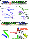
Figure 1
Figure 1
Structure-derived redefinition of a TALE repeat. (A) The traditional demarcation of a TALE repeat. Shown here is the secondary structure of a representative repeat in dHax3 (Deng et al., 2012a). RVD is indicated by red XX. Z stands for Glu or Gln. Please refer to Fig. S1B for the three-dimensional structure of a previously defined TALE repeat. (B) Re-demarcation of the TALE repeat allows the existence of integral number of repeats in TAL effectors. The structural elements are labeled with both new and previous (in bracket) designations. According to the new demarcation, the previous Helix a in repeat 11.5, which is identical to Helix a in any other repeat of dHax3, becomes Helix 12S of repeat 12. Similarly, the previous Helix 1b becomes Helix 2L. Within the new system, the residues in a TALE repeat are also re-numbered. The corresponding numbers within the previous system are bracketed. (C) The last helix of the N-domain in dHax3 is structurally similar to Helix b in the previously defined TALE repeat. The N-domain and the following Helix 1a (now Helix 1S) of dHax3 are colored green and cyan, respectively. Notably, the structural segment containing the last helix of the N-domain and Helix 1S can be reasonably well superimposed to the structural motif shown in Fig. 1B. Therefore, we define the last helix of the N-domain as Helix 1L of the first repeat in TAL effectors. (D) Structure-suggested re-demarcation of a TALE repeat. Based on the structures of TALE proteins, we wish to propose a new numbering system for a TALE repeat which starts with the invariant residue Gly (originally Gly14) and concludes with the base-recognition residue (originally residue 13, the 2nd residue of RVD)
Figure 2
Figure 2
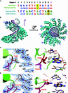
Structures of dHax3 variants in complex with their respective target DNA elements. (A) The sequences of the forward strand DNA and the corresponding RVDs used in the engineered dHax3 variants, which were designated dHax3-NI and dTALE, respectively. The RVDs that were not present in the reported dHax3 structure (Deng et al., 2012a) are shaded yellow. (B) Structural superimposition of DNA-bound dHax3 (grey) and dTALE (cyan). The two structures can be superimposed with an RMSD of 0.946 Å over 455 Cα atoms. The N-terminal domain of dTALE is colored blue. The PDB accession code for the DNA-bound dHax3 is 3V6T. (C and D) Structural basis for the recognition of bases A and G by Ile34 and Asn34. The 2Fo-Fc electron density map, shown in blue mesh, was contoured at 1.2 σ. The distances between the side group of Ile34 and base A are labeled in the unit of Å. The hydrogen bond between Asn34 and base G was indicated by red dashed line (lower right). All structure figures were prepared with PyMol (Schrodinger, 2010)
Figure 3
Figure 3
Structural basis for the recognition of bases adenine and guanine with new TALE codes. (A) A summary of the reported structures (Deng et al., ; Deng et al., 2012b) for the recognition of the indicated DNA bases by dHax3. Shown here are the 34th residues in the TALE repeats. (B and C) Structural basis for the recognition of bases A or G by a number of predicted or unpredicted TALE codes. Each panel represents a dHax3 variant in which the 34th residue in repeat 7 was replaced with the indicated amino acid. In total fifteen structures were determined (Tables S1–3). Notably, Asn34 and His34 each bind to A and G in different ways, which provides the structural basis for the distinctive recognition strengths revealed by genetic studies (Streubel et al., 2012)
Figure 4
Figure 4
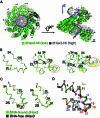
Structural basis underlying the structural plasticity of TALE repeats. (A) The structure of DNA-bound dHax3-NI determined at 2.2 Å (high) resolution exhibits distinct conformation from all the other DNA-bound dHax3 variants, including the same complex determined at 2.8 Å (low) resolution. The forward strands of DNA in the high and low resolution structures are colored pink and magenta, respectively. The two structures are superimposed against the 1st TALE repeat in dHax3. The N-terminal domain of the low resolution dHax3-NI is colored dark green. (B) Small structural variations in each repeat are amplified to prominent conformational changes seen in the overall structure in Fig. 4A. Shown here are the structural superimpositions against repeat 2 for one, two, and three repeats of the high (grey) and low (green) resolution structures of DNA-bound dHax3-NI. The segments that display the most pronounced structural changes are highlighted by orange circles. (C) Structural comparison of the 2nd repeat in the DNA-free (silver) and DNA-bound (green) dHax3 reveals that residues 14–22 display structural flexibility. Repeat 2 from the two structures are superimposed against either Helix S (left) or Helix L (right). (D) Structural comparison of 22 TALE repeats out of the high and low structures of dHax3-NI. For visual clarity, only Cα ribbons are shown. The residue numbers are colored from black to red with increasing deviations of the Cα atoms
Figure 5
Figure 5
Structural and functional analysis of the residues within a TALE repeat. (A) The first four residues (BBR, backbone binding residues) of a TALE repeat are responsible for DNA backbone binding. Left panel: DNA-bound dHax3 with the repeats relabeled according to the new demarcation defined in this manuscript. Only the backbone of the forward strand DNA is shown. Central panel: Gly1 and Gly2 in a TALE repeat bind to DNA phosphates through water-mediated H-bonds. Right panel: Lys3 and Gln4 coordinate the backbone phosphate of the forward strand DNA through direct or water-mediated H-bonds. Water molecules are shown as red spheres. H-bonds are represented as red dashed-lines. (B) Loop-stabilizing residue, His or Asn, at position 33 provides H-bond donor to interact with the carbonyl oxygen in Helix S. (C) The intra- and inter-repeat contacts are mediated mainly through van der Waals interactions. Residues that mediate the intra-(left) and inter-(right) repeat contacts are shown as sticks. (D) Composition analysis of the residues in a TALE repeat. The residues shown here for each position is adopted from a statistics of 2023 TALE repeats (Boch and Bonas, 2010). The residues that are involved in intra- or inter-repeat contacts are indicated by the blue squares above and shaded in cyan. Note that few residues from the RF (repeat flexibility) segment, which is colored yellow, are involved in the structural stabilization

References

    1. Adams PD, Grosse-Kunstleve RW, Hung LW, Ioerger TR, McCoy AJ, Moriarty NW, Read RJ, Sacchettini JC, Sauter NK, Terwilliger TC. PHENIX: building new software for automated crystallographic structure determination. Acta Crystallogr. 2002;58:1948–1954. - PubMed
    1. Bai J, Choi SH, Ponciano G, Leung H, Leach JE. Xanthomonas oryzae pv. oryzae avirulence genes contribute differently and specifically to pathogen aggressiveness. Mol Plant Microbe Interact. 2000;13:1322–1329. doi: 10.1094/MPMI.2000.13.12.1322. - DOI - PubMed
    1. Beumer KJ, Trautman JK, Christian M, Dahlem TJ, Lake CM, Hawley RS, Grunwald DJ, Voytas DF, Carroll D. Comparing ZFNs and TALENs for gene targeting in Drosophila. G3 (Bethesda) 2013;3(10):1717–1725. doi: 10.1534/g3.113.007260. - DOI - PMC - PubMed
    1. Boch J, Bonas U. Xanthomonas AvrBs3 family-type III effectors: discovery and function. Annu Rev Phytopathol. 2010;48:419–436. doi: 10.1146/annurev-phyto-080508-081936. - DOI - PubMed
    1. Boch J, Scholze H, Schornack S, Landgraf A, Hahn S, Kay S, Lahaye T, Nickstadt A, Bonas U. Breaking the code of DNA binding specificity of TAL-type III effectors. Science. 2009;326:1509–1512. doi: 10.1126/science.1178811. - DOI - PubMed

Publication types