. 2014 Jul 30;5(14):5198-208.
doi: 10.18632/oncotarget.2263.

Structural basis of nSH2 regulation and lipid binding in PI3Kα

Affiliations

Structural basis of nSH2 regulation and lipid binding in PI3Kα

Michelle S Miller et al. Oncotarget. .

Abstract

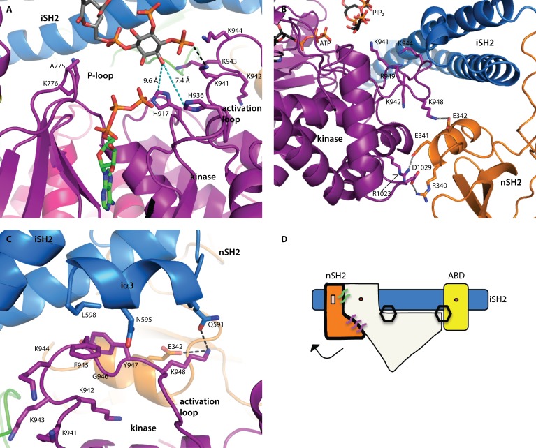
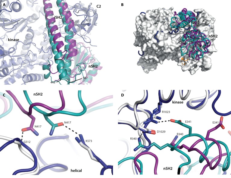
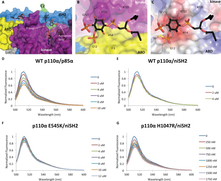
We report two crystal structures of the wild-type phosphatidylinositol 3-kinase α (PI3Kα) heterodimer refined to 2.9 Å and 3.4 Å resolution: the first as the free enzyme, the second in complex with the lipid substrate, diC4-PIP₂, respectively. The first structure shows key interactions of the N-terminal SH2 domain (nSH2) and iSH2 with the activation loop that suggest a mechanism by which the enzyme is inhibited in its basal state. In the second structure, the lipid substrate binds in a positively charged pocket adjacent to the ATP-binding site, bordered by the P-loop, the activation loop and the iSH2 domain. An additional lipid-binding site was identified at the interface of the ABD, iSH2 and kinase domains. The ability of PI3Kα to bind an additional PIP₂ molecule was confirmed in vitro by fluorescence quenching experiments. The crystal structures reveal key differences in the way the nSH2 domain interacts with wild-type p110α and with the oncogenic mutant p110αH1047R. Increased buried surface area and two unique salt-bridges observed only in the wild-type structure suggest tighter inhibition in the wild-type PI3Kα than in the oncogenic mutant. These differences may be partially responsible for the increased basal lipid kinase activity and increased membrane binding of the oncogenic mutant.


Conflict of interest statement

Under agreements between The Johns Hopkins University and various commercial entities, K.W.K. and B.V. are entitled to a share of the royalties received by The Johns Hopkins University on sales of products related to the diagnosis of mutant PIK3CA genes. The terms of these arrangements are being managed by The Johns Hopkins University in accordance with its conflict of interest policies.

Figures

Fig 1
Fig 1. Lipid-binding in PI3K
(A) Domain organization of the PI3Kα subunits, p110α and p85α, colored by domain. The positions of p110α hotspot oncogenic mutations are indicated. The niSH2 p85α construct used for crystallography is highlighted. (B) The substrate mimetic diC4-PIP2 binding site. This site is adjacent to the ATP binding site, between the activation loop and the P-loop of the kinase domain (shown in purple) and the iSH2 domain (shown in blue). (C) PI3Kα surface colored according to electrostatic potential, highlighting the positively charged PIP2 binding-site. (D) Activation loop sequence alignment, performed between the Class IA, II and III PI3Ks. Blue represents identical residues, orange represents similar residues, pink represents differences that have been identified as being important for PIP2 recognition and binding.
Fig 2
Fig 2. Structural insights into catalysis
All domains are colored according to the scheme in Fig. 1A. (A) The relationship between the two substrates was inferred by modeling a molecule of ATP into the binding site (from the alignment with the p110γ-ATP complex structure, PDB ID 1E8X). The 3’-hydroxyl is oriented toward the ATP γ-phosphate. There are two histidine residues in the binding site, which may deprotonate the 3’-hydroxyl for catalysis. Distances are shown in cyan colored dashed lines. (B) The nSH2 domain locks the activation loop in an inactive conformation via a salt-bridge between K948 (p110) and E342 (p85α). (C) The C-terminal residues of the iSH2 (p85α residues 587-602) form a short helix (iα3) which forms an interface with the activation loop. A hydrophobic stacking interaction is made between F945 (p110α) and L598 (p85α). Two key interactions between p85α (E342 and N591) are made with K948 of the activation loop, locking it in an inactive conformation. (D) Schematic representation of the p110α/niSH2 heterodimer showing the general position of the hydrogen bond network that locks p110α in an inactive conformation. In this scheme, p110α is represented in white. The two hexagons represent the PIP2 binding sites. The binding of phosphotyrosine residues at the helical-nSH2 interface causes a conformational change that breaks interactions with the activation loop, thereby activating the enzyme. In the basal state, this interface is maintained by key hydrogen bonds or salt-bridges between the subunits, represented by the purple and green lines.
Fig 3
Fig 3. Wild-type p110α has more interactions with the nSH2 domain than the oncogenic mutant H1047R
Superposition of the wild-type crystal structure p110α/niSH2 (PDB ID 4OVU) with the p110αH1047R/niSH2 oncogenic mutant (PDB ID 3HHM), obtained by aligning the two p110α molecules. The wild-type p110 and p85α are shown in dark blue and teal, respectively, while the p110αH1047R/niSH2 mutant structure is displayed as light grey (p110α) and purple (p85α). (A) The largest difference between the iSH2 domains is highlighted with a dashed line, measured between the Cα atoms of p85α 450 in each structure. (B) The 14° rotation of the nSH2 domain is identified with an orange arrow. p110αH1047R is shown as a surface representation. (C) Differences in the interactions between the helical and nSH2 domains of the wild-type and oncogenic mutant structures. (D) Two key salt-bridges between the kinase domain and nSH2 are present in the wild-type but absent in the oncogenic mutant structure.
Fig 4
Fig 4. Two PIP2 molecules bound to p110α/niSH2
p110α/niSH2 in complex with diC4-PIP2 is shown as a molecular surface with the kinase domain colored in purple, ABD domain in yellow, helical domain in pink, C2 in green, iSH2 in blue and nSH2 in orange. PIP2 molecules are shown as sticks with grey carbons. (A) Two molecules of PIP2 bind at the interface between p110α and iSH2 of p85α. The distance between the two binding sites is ~21 Å. (B) A second molecule of PIP2 binds at the interface between the ABD (yellow) and kinase (purple) domains. (C) The surface of the second PIP2 binding site colored according to the electrostatic potential shows a very hydrophobic surface, suggesting possibly a general lipid-binding site rather than a specific PIP2 binding site. (Fig. 4D-G) PI3K clusters PIP2 in model membrane vesicles containing 50 nM of BODIPY®-FL-PIP2. The highest normalized emission intensity corresponds to the vesicles alone. Each subsequent spectrum represents an incremental addition of the corresponding protein. All experiments were performed with N=3. Graphs shown are representative and present the data from a single experiment. (D) Wild-type p110α/p85α quenches 20% of the signal at 4 μM. (E) Wild-type p110α/niSH2 quenches in a similar fashion to the full-length complex. (F) The displayed quenching by p110αE545K/niSH2 is similar to wild-type p110α/niSH2. (G) p110αH1047R/niSH2 quenches the signal with a much higher potency than wild-type. Only 500 nM of protein is required to quench the signal by 20%.

References

    1. Fyffe C, Buus R, Falasca M. Genetic and Epigenetic Regulation of Phosphoinositide 3-kinase Isoforms. Curr Pharm Des. 2013;19:680–6. - PubMed
    1. Broderick DK, Di C, Parrett TJ, Samuels YR, Cummins JM, McLendon RE, Fults DW, Velculescu VE, Bigner DD, Yan H. Mutations of PIK3CA in Anaplastic Oligodendrogliomas, High-Grade Astrocytomas, and Medulloblastomas. Cancer Res. 2004;64:5048–50. - PubMed
    1. Campbell IG, Russell SE, Choong DYH, Montgomery KG, Ciavarella ML, Hooi CSF, Cristiano BE, Pearson RB, Phillips WA. Mutation of the PIK3CA gene in ovarian and breast cancer. Cancer Res. 2004;64:7678–81. - PubMed
    1. Levine DA, Bogomolniy F, Yee CJ, Lash A, Barakat RR, Borgen PI, Boyd J. Frequent mutation of the PIK3CA gene in ovarian and breast cancers. Clin Cancer Res. 2005;11:2875–8. - PubMed
    1. Bachman KE, Argani P, Samuels Y, Silliman N, Ptak J, Szabo S, Konishi H, Karakas B, Blair BG, Lin C, Peters BA, Velculescu VE, Park BH. The PIK3CA gene is mutated with high frequency in human breast cancers. Cancer Biol Ther. 2004;3:772–5. - PubMed

Publication types

Substances

LinkOut - more resources