. 2015 Jul 3;290(27):16539-49.
doi: 10.1074/jbc.M114.622928. Epub 2015 May 14.

Probing the Solution Structure of IκB Kinase (IKK) Subunit γ and Its Interaction with Kaposi Sarcoma-associated Herpes Virus Flice-interacting Protein and IKK Subunit β by EPR Spectroscopy

Affiliations

Probing the Solution Structure of IκB Kinase (IKK) Subunit γ and Its Interaction with Kaposi Sarcoma-associated Herpes Virus Flice-interacting Protein and IKK Subunit β by EPR Spectroscopy

Claire Bagnéris et al. J Biol Chem. .

Erratum in

Abstract

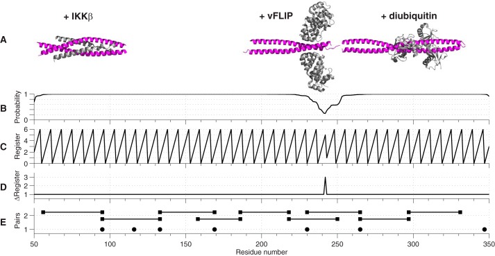
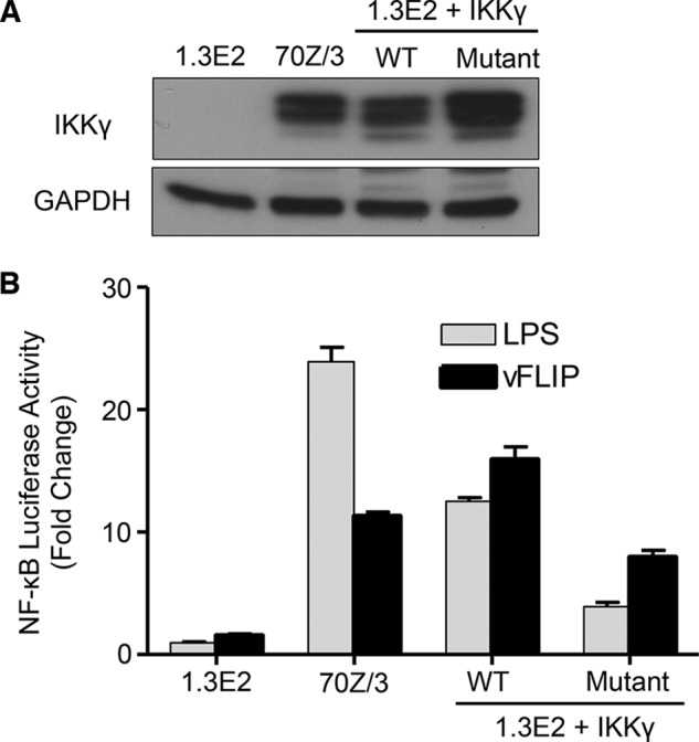
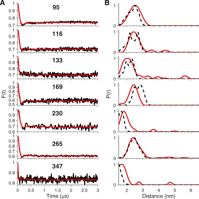
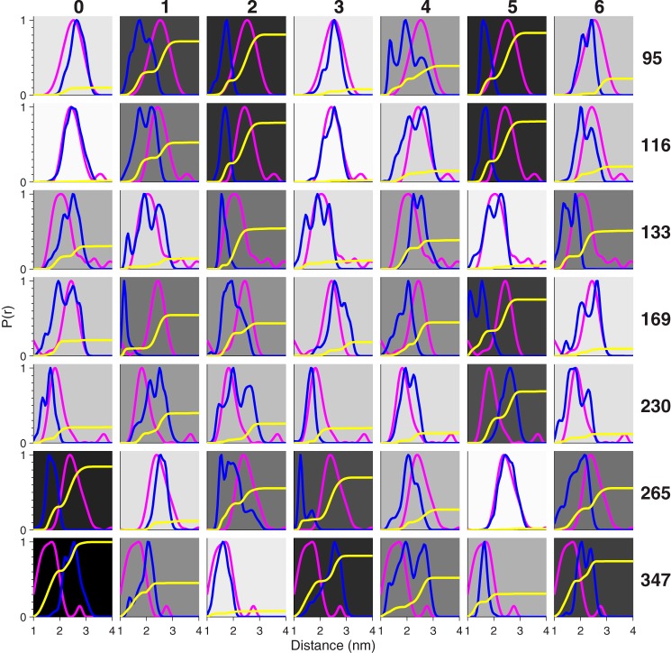
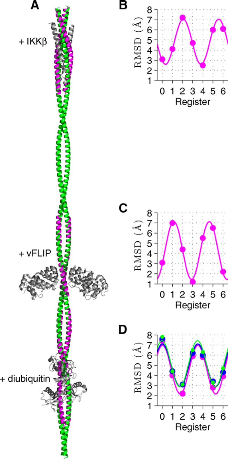
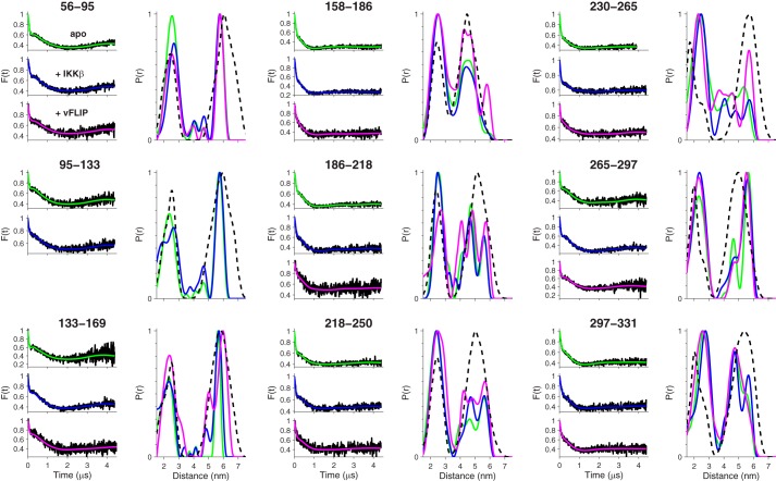
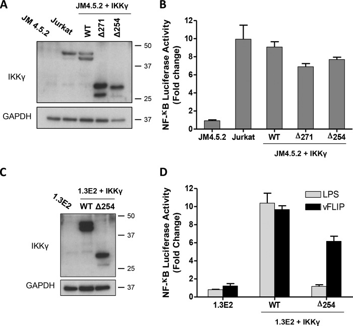
Viral flice-interacting protein (vFLIP), encoded by the oncogenic Kaposi sarcoma-associated herpes virus (KSHV), constitutively activates the canonical nuclear factor κ-light-chain-enhancer of activated B cells (NF-κB) pathway. This is achieved through subversion of the IκB kinase (IKK) complex (or signalosome), which involves a physical interaction between vFLIP and the modulatory subunit IKKγ. Although this interaction has been examined both in vivo and in vitro, the mechanism by which vFLIP activates the kinase remains to be determined. Because IKKγ functions as a scaffold, recruiting both vFLIP and the IKKα/β subunits, it has been proposed that binding of vFLIP could trigger a structural rearrangement in IKKγ conducive to activation. To investigate this hypothesis we engineered a series of mutants along the length of the IKKγ molecule that could be individually modified with nitroxide spin labels. Subsequent distance measurements using electron paramagnetic resonance spectroscopy combined with molecular modeling and molecular dynamics simulations revealed that IKKγ is a parallel coiled-coil whose response to binding of vFLIP or IKKβ is localized twisting/stiffening and not large-scale rearrangements. The coiled-coil comprises N- and C-terminal regions with distinct registers accommodated by a twist: this structural motif is exploited by vFLIP, allowing it to bind and subsequently activate the NF-κB pathway. In vivo assays confirm that NF-κB activation by vFLIP only requires the N-terminal region up to the transition between the registers, which is located directly C-terminal of the vFLIP binding site.

Keywords: IKKγ; NEMO; NF-kappa B (NF-κB); electron paramagnetic resonance (EPR); molecular dynamics; molecular modeling; protein structure; signalosome; vFLIP; virology.


Figures

FIGURE 1.
FIGURE 1.
SDS-PAGE Coomassie-stained gels of the final size-exclusion profiles obtained for: IKKγ; IKKγ·vFLIP complex; IKKγ·IKKβ complex; IKKγ·vFLIP·IKKβ ternary complex of the 56–95 double mutant.
FIGURE 2.
FIGURE 2.
Diagram illustrating how the register of the coiled-coil translates into distance distributions between two pairs of nitroxide spin labels. Limiting cases in which (A) all 4 labels are in a plane or (B) the two vectors joining the pairs are perpendicular. C, geometry is constrained by the intra-pair distance (designated a), the inter-pair distance (designated b) and dihedral angle (designated θ) between the vectors joining the pairs. D, illustration of the variation of the distance distribution for two pairs of labels as a function of θ for a = 2.5 nm and b = 5 nm with Gaussian linewidth of 0.2 nm.
FIGURE 3.
FIGURE 3.
A, crystal structures of fragments of IKKγ (magenta) in complex with IKKβ, vFLIP, and diubiuitin (gray) aligned according to their position in the sequence. B, probability of a region adopting a coiled-coil arrangement based on a window of 28 residues predicted from the primary sequence of IKKγ. C, predicted position of residues within a coiled-coil based on the heptameric repeat of the motif: each residue has a predicted position from 0–6 and a regular zigzag pattern implies a perfect coiled-coil. D, increment of the register of a residue with respect to its predecessor. An increment of +1 implies a perfect coiled-coil. E, positions of nitroxide spin labels for singly labeled IKKγ (solid circles) and doubly labeled IKKγ (solid squares joined by solid lines).
FIGURE 4.
FIGURE 4.
NF-κB reporter assays for the vFLIP mutant in which all 5 out of 6 cysteines were mutated to serine. A, Western blot analysis of IKKγ expression level in IKKγ knock-out mouse PreB cell line 1.3E2, respective parental cells 70Z/3 and IKKγ wild type or mutant reconstituted 1.3E2 cells. B, NF-κB activation was measured in the above-mentioned cell lines using the BrightGlo Luciferase assay system, 6 h after stimulation with LPS (10 μg ml−1) or 48 h after transduction with KSHV vFLIP LV (MOI = 50). Bars represent mean fold induction values ± S.D.
FIGURE 5.
FIGURE 5.
EPR distance measurement on singly-labeled IKKγ. A, background-corrected dipolar evolution (black lines) and their fits (red lines). B, distance distributions (red lines) derived from the data in A and predicted distance distributions (black dashed lines) derived from the static 3/5 hybrid model.
FIGURE 6.
FIGURE 6.
Validation of the model structures. Comparison of the distance distributions obtained from the EPR experiments for the singly-labeled IKKγ (magenta lines) with the predicted distance distributions (blue lines) using a rotamer library approach for the 0–6 register models. The cumulative sum of the squares of the differences between the experimental and predicted distributions is used to provide a metric for goodness of fit (yellow lines). The background has been gray-shaded according to the relative final amplitude of the yellow lines: a white background implies perfect match, whereas a black background implies poor agreement.
FIGURE 7.
FIGURE 7.
Comparison of the 3/5 model of IKKγ with crystal structures. A, 3/5 model (green) is overlaid with crystal structures of IKKγ fragments (magenta) with IKKβ, vFLIP, and diubiquitin (gray). Plots of the RMSDs of the positions of the non-hydrogenic atoms in the coiled-coil model structures (Registers 0–6) compared with those in the known crystal structures: (B) IKKγ + IKKβ (22); (C) IKKγ + vFLIP (12); (D) IKKγ ubiquitin binding region: apo (23) (blue), with diubiquitin (24) (magenta), and with Hiop (25) (green). The curves are fits to the RMSDs assuming a sinusoidal pattern that completes two oscillations within the heptad repeat.
FIGURE 8.
FIGURE 8.
EPR distance measurements on doubly-labeled IKKγ, apo and complexed with IKKβ and vFLIP. Fits to the baseline-corrected dipolar evolution and corresponding distance distributions for apo IKKγ (green lines) and IKKγ complexed with IKKβ (blue lines) and vFLIP (magenta lines). The distance distributions for the doubly labeled IKKγ derived from the coarse-grained MD simulation of the 3/5 hybrid model are shown as a dashed black line.
FIGURE 9.
FIGURE 9.
NF-κB luciferase reporter assays for the IKKγ Δ254–419 and Δ271–419 truncation mutants and D242R mutant. A, Western blot analysis of IKKγ expression level in IKKγ knock-out human T-cells JM4.5.2, parental Jurkat cells and IKKγ wild type or mutant reconstituted JM4.5.2 cells. B, NF-κB activation was measured using the BrightGlo Luciferase assay system 48 h after transduction with KSHV vFLIP LV (MOI = 50). Bars represent mean fold induction values ± S.D. C, Western blot analysis of IKKγ expression levels in IKKγ knock-out mouse PreB cell line 1.3E2, parental cells 70Z/3, and 1.3E2 cells reconstituted with IKKγ wild type; Δ254–419 mutant or IKKγ D242R. D, NF-κB activation was measured using the BrightGlo Luciferase assay system, 6 h after stimulation with LPS (10 μg ml−1) or 48 h after transduction with KSHV vFLIP LV. Bars represent mean of relative luminescence unit (RLU) values ± S.D.

References

    1. Scheidereit C. (2006) IκB kinase complexes: gateways to NF-κB activation and transcription. Oncogene 25, 6685–6705 - PubMed
    1. DiDonato J. A., Hayakawa M., Rothwarf D. M., Zandi E., Karin M. (1997) A cytokine-responsive IκB kinase that activates the transcription factor NF-κB. Nature 388, 548–554 - PubMed
    1. Mercurio F., Zhu H., Murray B. W., Shevchenko A., Bennett B. L., Li J., Young D. B., Barbosa M., Mann M., Manning A., Rao A. (1997) IKK-1 and IKK-2: cytokine-activated IκB kinases essential for NF-κB activation. Science 278, 860–866 - PubMed
    1. Israël A. (2010) The IKK complex, a central regulator of NF-κB activation. Cold Spring Harb. Perspect. Biol. 2, a000158. - PMC - PubMed
    1. Hiscott J., Nguyen T. L., Arguello M., Nakhaei P., Paz S. (2006) Manipulation of the NF-κB pathway and the innate immune response by viruses. Oncogene 25, 6844–6867 - PMC - PubMed

Publication types

Substances