. 2014 Sep 5;345(6201):1188-91.
doi: 10.1126/science.1255030.

How the ribosome hands the A-site tRNA to the P site during EF-G-catalyzed translocation

Affiliations

How the ribosome hands the A-site tRNA to the P site during EF-G-catalyzed translocation

Jie Zhou et al. Science. .

Abstract

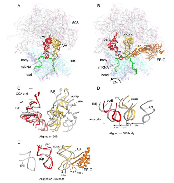
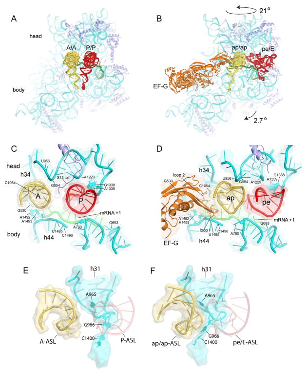
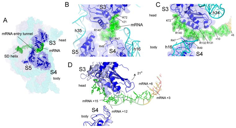
Coupled translocation of messenger RNA and transfer RNA (tRNA) through the ribosome, a process catalyzed by elongation factor EF-G, is a crucial step in protein synthesis. The crystal structure of a bacterial translocation complex describes the binding states of two tRNAs trapped in mid-translocation. The deacylated P-site tRNA has moved into a partly translocated pe/E chimeric hybrid state. The anticodon stem-loop of the A-site tRNA is captured in transition toward the 30S P site, while its 3' acceptor end contacts both the A and P loops of the 50S subunit, forming an ap/ap chimeric hybrid state. The structure shows how features of ribosomal RNA rearrange to hand off the A-site tRNA to the P site, revealing an active role for ribosomal RNA in the translocation process.


Figures

Figure 1
Figure 1. Structure of trapped translocation intermediate containing EF-G, mRNA, and two partially-translocated tRNAs
(A) 70S ribosome with tRNAs bound in classical A/A and P/P states (17); (B) Translocation intermediate complex, showing tRNAs trapped in intermediate chimeric hybrid ap/ap and pe/E states. (C) Comparison of positions of tRNA in classical states and in the translocation intermediate, aligned on the 50S subunit; (D) relative positions of tRNA ASLs, aligned on the 30S subunit body or (E) head. Molecular components are colored throughout as: 16S rRNA, cyan; 30S proteins, blue; 23S rRNA, grey; 5S rRNA, light blue; 50S proteins, magenta; mRNA, green; A/A and ap/ap tRNAs, yellow; P/P and pe/E tRNAs, red; EF-G, orange.
Figure 2
Figure 2. Movement of tRNA ASLs on the 30S subunit, and capture of translocating A-tRNA by P-site elements of the 30S subunit head
(A–B) Positions of tRNAs in the (A) classical-state ribosome and (B) translocation intermediate. (C–D) Interactions of the tRNA ASLs and mRNA as they move from the (C) A and P classical states to the (D) ap and pe chimeric hybrid states. (E–F) Rearrangement of the 966 loop (h31) of 16S rRNA in the 30S subunit head from its (E) classical P-tRNA-binding position to (F) forms a surface-fitting pocket around the ap/ap tRNA ASL.
Figure 3
Figure 3. Contact between the downstream region of mRNA with the head of the 30S subunit
(A) The entry to the downstream mRNA entry tunnel is surrounded by proteins uS3, uS4, and uS5. Positions +14 to +21 contact a positively charged patch on the surface of protein uS3 in the 30S head (Fig. S17). (B) Path of the downstream mRNA and (C) 90° rotated view. (D) Docking on the 30S body of the classical structure (17) (gray) shows that rotation of the head in the ap/ap intermediate structure translocates the mRNA by one codon. A-codon (yellow) and P-codon (red).
Figure 4
Figure 4. The acceptor end of ap/ap tRNA simultaneously contacts the A and P loops of 23S rRNA
During translocation, the 3′ acceptor end of peptidyl-tRNA moves from the (A) classical A/A state to the (B) intermediate ap/ap state to the (C) classical P/P states, facilitated by conformational changes in the A and P loops.

References

    1. Rodnina MV, Savelsbergh A, Katunin VI, Wintermeyer W. Nature. 1997;385:37–41. - PubMed
    1. Moazed D, Noller HF. Nature. 1989;342:142–148. - PubMed
    1. Frank J, Agrawal RK. Nature. 2000;406:318–322. - PubMed
    1. Valle M, et al. Cell. 2003;114:123–134. - PubMed
    1. Agirrezabala X, et al. Mol Cell. 2008;32:190–197. - PMC - PubMed

Publication types

MeSH terms