. 2014 Sep 25;8(6):1668-1676.
doi: 10.1016/j.celrep.2014.08.010. Epub 2014 Sep 4.

Binding-pocket and lid-region substitutions render human STING sensitive to the species-specific drug DMXAA

Affiliations

Binding-pocket and lid-region substitutions render human STING sensitive to the species-specific drug DMXAA

Pu Gao et al. Cell Rep. .

Abstract

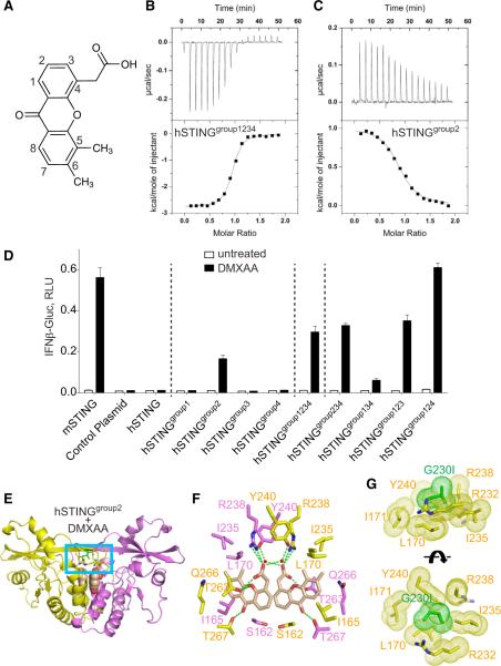
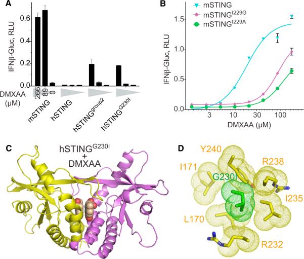
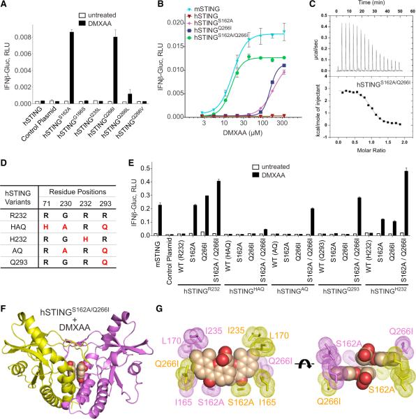
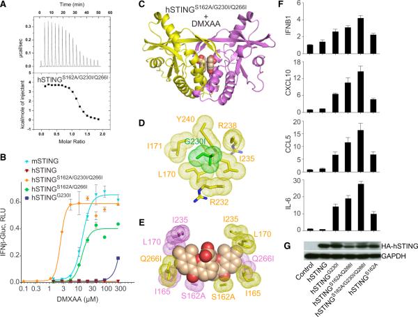
The drug DMXAA (5,6-dimethylxanthenone-4-acetic acid) showed therapeutic promise against solid tumors in mouse models but subsequently failed in human clinical trials. DMXAA was later discovered to activate mouse, but not human, STING, an adaptor protein in the cyclic dinucleotide cGAMP-mediated signaling pathway, inducing type I interferon expression. To facilitate the development of compounds that target human STING, we combined structural, biophysical, and cellular assays to study mouse and human chimeric proteins and their interaction with DMXAA. We identified a single substitution (G230I) that enables a DMXAA-induced conformational transition of hSTING from an inactive "open" to an active "closed" state. We also identified a substitution within the binding pocket (Q266I) that cooperates with G230I and the previously identified S162A binding-pocket point substitution, rendering hSTING highly sensitive to DMXAA. These findings should facilitate the reciprocal engineering of DMXAA analogs that bind and stimulate wild-type hSTING and their exploitation for vaccine-adjuvant and anticancer drug development.


Figures

Figure 1
Figure 1. Replacement of Nonconserved Residues of hSTING with Its Murine Counterparts Enables Recognition of DMXAA as well as the Crystal Structure of DMXAA Bound to hSTINGgroup2
(A) Chemical formula of DMXAA. (B and C) ITC binding curves for complex formation between DMXAA bound to hSTINGgroup1234 (aa 140–379) (B) and hSTINGgroup2 (C). (D) 293T cells were transfected with IFN-β reporter constructs and STING variants as indicated. At 12 hr after transfection, cells were stimulated with 0.18 mM DMXAA (50 μg/ml). Luciferase activity was determined after another 12 hr. Dotted lines separate (from left to right) WT controls, single group mutants, hSTINGgroup1234, and triple-group mutants. Shown are raw values of Gaussian luciferase activity normalized to constitutive Firefly luciferase values. Values depicted are the means of triplicates + SEM and are representative of three independent experiments. (E) The 1.88Å crystal structure of DMXAA bound to hSTINGgroup2 (aa 155–341). The symmetrical hSTINGgroup2 dimer is shown in a ribbon representation, with individual monomers colored in yellow and magenta. The DMXAA (in a space-filling representation) is bound in the central cavity at the interface between the two monomers. (F) Intermolecular contacts in the complex. The bound DMXAA is shown in biscuit color, with individual STING subunits in the symmetrical dimer shown in yellow and magenta. (G) Two expanded views of the hydrophobic interactions of the G230I substitution (in green) in the complex (blue box region in E). Other residues lining the hydrophobic pocket are shown in yellow. See also Figures S1 and S2.
Figure 2
Figure 2. G230 in hSTING and I229 in mSTING Are Essential for DMXAA Species Selectivity
(A) 293T cells were transfected with IFN-β reporter constructs and STING variants as indicated. At 12 hr after transfection, cells were stimulated with ascending concentrations of DMXAA. Luciferase activity was determined after another 12 hr. Shown are the means of triplicates + SEM, representative of three independent experiments. (B) 293T cells were transfected with mSTING variants and reporter constructs. Stimulation and luciferase assay were performed as described in (A). Dose responses are representative of two independent experiments. (C) The 2.51Å crystal structure of DMXAA bound to hSTINGG230I (aa 155–341). The representations and color codes are the same as in Figure 1E. (D) Detailed hydrophobic interactions in the complex of DMXAA bound to hSTINGG230I, with the same representations and color codes as in Figure 1G. See also Figure S3.
Figure 3
Figure 3. S162A and Q266I Substitutions Render hSTING Sensitive to DMXAA
(A) 293T cells were transfected with reporter constructs and the indicated hSTING variants. At 12 hr after transfection, cells were stimulated with 0.18 mM DMXAA for another 12 hr, followed by luciferase assay. Shown are means of triplicates + SEM, representative of three independent experiments. (B) DMXAA dose-response curves of 293T cells transfected with the indicated STING variants, illustrating one representative experiment out of three independent experiments. (C) ITC binding curve for complex formation between DMXAA bound to hSTINGS162A/Q266I (aa 140–379). (D) Natural variants of hSTING (Yi et al., 2013). Five hSTING variants (shown in the left column) were studied in this work. The amino acid variations are shown in the right column and colored in red. (E) 293T cells were transfected and stimulated as in (A). Shown are the S162A and Q266I mutants of major hSTING alleles. WT denotes the respective allele in this context. Shown are the means of triplicates + SEM, representative of three independent experiments. (F) The 2.42Å crystal structure of DMXAA bound to hSTINGS162A/Q266I (aa 155–341). The representations and color codes are the same as used in Figure 1E. (G) Two alternate views of the hydrophobic interactions of DMXAA with hSTINGS162A/Q266I. The two bound DMXAA molecules are shown in space-filling representation, with surrounded hydrophobic side chains shown in stick and dot representations. See also Figures S1, S3, and S4.
Figure 4
Figure 4. Triple Substitution of G230I/S162A/Q266I Yields an hSTING Variant with Higher Affinity to DMXAA in Comparison to mSTING and Robust Stimulation of Cytokines and Chemokines in Mouse Cells
(A) ITC binding curve for complex formation between DMXAA bound to hSTINGS162A/G230I/Q266I (aa 140–379). (B) 293T cells were transfected with reporter constructs and the indicated STING variants. After 12 hr, cells were stimulated with increasing concentrations of DMXAA for another 12 hr, followed by luciferase assay. Individual data points are means of triplicates ± SEM. Representative of three independent experiments. (C) The 2.37Å crystal structure of DMXAA bound to hSTINGS162A/G230I/Q266I (aa 155–341). The representations and color codes are the same as in Figure 1E. (D) Hydrophobic interactions of G230I substitution in the complex of DMXAA bound to hSTINGS162A/G230I/Q266I. The representations and color codes are the same as in Figure 1G. (E) Hydrophobic interactions of DMXAA in the ligand binding pocket, with the same representations and color codes as in Figure 3G. (F) BMDCs (1 × 10⁶) generated from a STINGGt/Gt mouse were transduced with retroviruses expressing WT and various hSTING mutants. Two days after viral infection, the BMDCs were treated with 50 mg/ml of DMXAA for 3 hr and cells were collected for real-time PCR to measure IFNB1, CXCL10, CCL5, and IL-6 mRNA levels. Data shown are means ± SEM (n = 3), representative of two independent experiments. (G) BMDCs were transduced with retroviruses expressing WT hSTING and various hSTING mutants. Cells were collected 2 days after retroviral infection and the levels of hSTING were determined by western blot analysis. See also Figure S4.

References

    1. Ablasser A, Goldeck M, Cavlar T, Deimling T, Witte G, Röhl I, Hopfner KP, Ludwig J, Hornung V. cGAS produces a 2′-5′-linked cyclic dinucleotide second messenger that activates STING. Nature. 2013;498:380–384. - PMC - PubMed
    1. Baguley BC, Ching LM. DMXAA: an antivascular agent with multiple host responses. Int. J. Radiat. Oncol. Biol. Phys. 2002;54:1503–1511. - PubMed
    1. Burdette DL, Vance RE. STING and the innate immune response to nucleic acids in the cytosol. Nat. Immunol. 2013;14:19–26. - PubMed
    1. Burdette DL, Monroe KM, Sotelo-Troha K, Iwig JS, Eckert B, Hyodo M, Hayakawa Y, Vance RE. STING is a direct innate immune sensor of cyclic di-GMP. Nature. 2011;478:515–518. - PMC - PubMed
    1. Conlon J, Burdette DL, Sharma S, Bhat N, Thompson M, Jiang Z, Rathinam VAK, Monks B, Jin T, Xiao TS, et al. Mouse, but not human STING, binds and signals in response to the vascular disrupting agent 5,6-dimethylxanthenone-4-acetic acid. J. Immunol. 2013;190:5216–5225. - PMC - PubMed

Publication types

MeSH terms