. 2015 Feb 6;290(6):3198-208.
doi: 10.1074/jbc.M114.624536. Epub 2014 Dec 9.

Crystal structures of enterovirus 71 (EV71) recombinant virus particles provide insights into vaccine design

Affiliations

Crystal structures of enterovirus 71 (EV71) recombinant virus particles provide insights into vaccine design

Ke Lyu et al. J Biol Chem. .

Abstract

Hand-foot-and-mouth disease (HFMD) remains a major health concern in the Asia-Pacific regions, and its major causative agents include human enterovirus 71 (EV71) and coxsackievirus A16. A desirable vaccine against HFMD would be multivalent and able to elicit protective responses against multiple HFMD causative agents. Previously, we have demonstrated that a thermostable recombinant EV71 vaccine candidate can be produced by the insertion of a foreign peptide into the BC loop of VP1 without affecting viral replication. Here we present crystal structures of two different naturally occurring empty particles, one from a clinical C4 strain EV71 and the other from its recombinant virus containing an insertion in the VP1 BC loop. Crystal structure analysis demonstrated that the inserted foreign peptide is well exposed on the particle surface without significant structural changes in the capsid. Importantly, such insertions do not seem to affect the virus uncoating process as illustrated by the conformational similarity between an uncoating intermediate of another recombinant virus and that of EV71. Especially, at least 18 residues from the N terminus of VP1 are transiently externalized. Altogether, our study provides insights into vaccine development against HFMD.

Keywords: Crystal Structure; Infectious Disease; Vaccine; Vaccine Development; Virus Structure.


Figures

FIGURE 1.
FIGURE 1.
Design of EV71 insertion mutants. A, top view of a pentameric structure of EV71 with residues 97–105 of VP1 colored in red. This region constitutes a neutralizing epitope and is well displayed on the virus surface. B, EV71 genome organization and design of EV71 insertion mutants. In the case of EV71-NE1, an insertion of 8 amino acids resulted in the loss of residues 101 and 102 (TN) in VP1 in the rescued mutant virus.
FIGURE 2.
FIGURE 2.
Purification and characterization of empty particles and virions from EV71-WT and insertion mutants. A, purification of EV71. The protein composition of the purified particles obtained from discontinuous sucrose gradient ultracentrifugation was analyzed by 12% SDS-PAGE. Lane M, molecular weight marker; lanes 10–14, empty particles; lane 15, virions. The empty particle is composed of three structural proteins, VP0, VP1, and VP3; the virion is composed of VP1, VP2 (VP0 cleavage product), VP3, and VP4 (not visible on this gel). The calculated molecular masses of VP1, VP2, VP3, and VP4 are 32.6, 27.7, 26.4, and 7.5 kDa, respectively. B, purified EV71-NE1 analyzed by SDS-PAGE. Lane 1, empty particles of EV71-NE1; lane 2, virions of EV71-NE1; lane 3, empty particles and virions of EV71. C, purified EV71-N6 analyzed by SDS-PAGE. Lane 1, empty particles of EV71-N6; lane E, empty particles of EV71; lane 2, virions of EV71-N6. D, a transmission EM image of EV71-NE1 empty particles. E, a transmission EM image of EV71-NE1 virions. Scale bar, 100 nm.
FIGURE 3.
FIGURE 3.
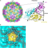
Structure of EV71 empty particles. A, left, a schematic representation of the EV71 empty particle viewed along the 2-fold axis with VP1, VP0, and VP3 colored in magenta, yellow, and cyan, respectively. Right, a schematic representation of a protomer with the positions of the icosahedral symmetry elements indicated. B, a hydrogen bonding network at the 5-fold axis channel. The amino group in the side chain of Lys-182 (colored black) interacts with Asp-185 (colored red) of a neighboring VP1 through hydrogen bonds.
FIGURE 4.
FIGURE 4.
Structures of empty particles from EV71 insertion mutants. A, surface representation of the EV71-NE1 empty particle viewed along the 2-fold axis. EV71 capsid proteins VP1, VP0, and VP3 are colored in magenta, yellow, and cyan, respectively. The inserted amino acids are colored in blue. B, superposition of VP1 from EV71 and EV71-NE1 empty particles. Residues 73–296 are modeled in the EV71 empty particle and colored in gray. Residues 72–303 are modeled in the EV71-NE1 empty particle and colored in red. C, superimposition of VP0 (VP2) from EV71 and EV71-NE1 empty particles. The color scheme is the same as in B. Residues 82–319 (VP2, 13–250) are modeled in the EV71 empty particle. Residues 80–321 (VP2, 11–252) are modeled in the EV71-NE1 empty particle. D, superimposition of VP3 from EV71 and EV71-NE1 empty particle. The proteins are colored the same as in B. Residues 1–175 and 189–238 are modeled in the EV71 empty particle. Residues 1–178 and 189–240 are modeled in the EV71-NE1 empty particle. E, the electron densities corresponding to the inserted NE1 peptide. AA, amino acids.
FIGURE 5.
FIGURE 5.
Surface representation of the EV71-NE1 virion structure by homology modeling. The capsid proteins VP1–VP4 are colored in magenta, yellow, cyan, and red, respectively. The inserted peptide (NE1) is colored in blue. Homology modeling showed that all 60 copies of the NE1 peptide are uniformly displayed on the surface of the EV71-NE1 virion.
FIGURE 6.
FIGURE 6.
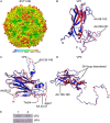
Structural comparisons between the uncoating intermediates of EV71 and EV71-N6. A, radius-colored surface representation of the EV71-N6 uncoating intermediate viewed along the 2-fold axis. The surface is colored from blue to red according to the distance from the particle center (blue represents the closest). B, superposition of VP1. Residues 73–98 and 119–313 of VP1 in EV71-N6 are colored in red, whereas residues 72–296 of VP1 in EV71 are colored in blue. C, superposition of VP2. Residues 11–47 and 54–252 of VP2 in EV71-N6 are colored in red, whereas residues 16–47 and 54–250 in EV71 are colored in blue. D, superposition of VP3. Residues 1–174 and 190–238 of VP3 in EV71-N6 are colored in red, whereas residues 1–175 and 189–236 in EV71 are colored in blue. E, detection of VP4 in the EV71-N6 uncoating intermediate after trypsin digestion. The uncoating intermediate was treated by trypsin digestion as described under “Experimental Procedures.” The digested samples were analyzed by Western blotting using anti-VP4 antibodies with VP2 as an internal control. Lane 1, hanging drops containing crystals of the uncoating intermediate (1 μg) incubated at 16 °C for 1 h; lane 2, hanging drops containing crystals of the uncoating intermediate (1 μg) digested with trypsin at 16 °C for 1 h. VP4 in the uncoating intermediate was resistant to protease digestion. AA, amino acids.
FIGURE 7.
FIGURE 7.
VP0 and the N-terminal regions of VP1 are at least transiently exposed. A, the empty particle junction is surrounded by VP1 (magenta), VP0 (yellow), and VP3 (cyan). VP1 from a neighboring protomer is colored in red. The visible N terminus of VP1 is located at the base of the junction (from the top view). B, a series of truncated products (VP1-a, VP1-b, VP1-c, and VP1-d) were detected with anti-VP1 antibodies. Lane 1, empty particles (1 μg) incubated at 16 °C for 1 h. Lane 2, empty particles (1 μg) digested by trypsin at 16 °C for 1 h. VP1-a, VP1-b, and VP1-c were detected. Lane 3, empty particles (1 μg) incubated at 37 °C for 1 h. Lane 4, empty particles (1 μg) digested by trypsin at 37 °C for 1 h. VP1-a, VP1-b, VP1-c, and VP1-d were detected. The N-terminal amino acid sequence of VP1-a is “ALTHA” (residues 19–23 of VP1). C, a band smaller than VP0 (VP0-a) was detected with anti-VP4 antibodies. Lane 1, empty particles (1.6 μg) incubated at 16 °C for 1 h. Lane 2, empty particles (1.6 μg) digested by trypsin at 16 °C for 1 h. VP0-a was detected. Lane 3, empty particles (1.6 μg) incubated at 37 °C for 1 h. Lane 4, empty particles (1.6 μg) digested by trypsin at 37 °C at 1 h. VP0-a was detected. D, the digestion profile of the empty particles is similar to that of the uncoating particles obtained by in vitro heating. Lane 1, uncoating particles (1 μg) incubated at 16 °C for 1 h. Lane 2, uncoating particles (1 μg) digested by trypsin at 16 °C for 1 h. VP1-a and VP1-b were detected. Lane 3, empty particles (1 μg) incubated at 16 °C for 1 h. Lane 4, empty particles (1 μg) digested by trypsin at 16 °C for 1 h. VP1-a and VP1-b were detected.

References

    1. McMinn P. C. (2012) Recent advances in the molecular epidemiology and control of human enterovirus 71 infection. Curr. Opin. Virol. 2, 199–205 - PubMed
    1. Wang G., Cao R. Y., Chen R., Mo L., Han J. F., Wang X., Xu X., Jiang T., Deng Y. Q., Lyu K., Zhu S. Y., Qin E. D., Tang R., Qin C. F. (2013) Rational design of thermostable vaccines by engineered peptide-induced virus self-biomineralization under physiological conditions. Proc. Natl. Acad. Sci. U.S.A. 110, 7619–7624 - PMC - PubMed
    1. Martin A., Wychowski C., Couderc T., Crainic R., Hogle J., Girard M. (1988) Engineering a poliovirus type 2 antigenic site on a type 1 capsid results in a chimaeric virus which is neurovirulent for mice. EMBO J. 7, 2839–2847 - PMC - PubMed
    1. Murray M. G., Kuhn R. J., Arita M., Kawamura N., Nomoto A., Wimmer E. (1988) Poliovirus type 1/type 3 antigenic hybrid virus constructed in vitro elicits type 1 and type 3 neutralizing antibodies in rabbits and monkeys. Proc. Natl. Acad. Sci. U.S.A. 85, 3203–3207 - PMC - PubMed
    1. Evans D. J., McKeating J., Meredith J. M., Burke K. L., Katrak K., John A., Ferguson M., Minor P. D., Weiss R. A., Almond J. W. (1989) An engineered poliovirus chimaera elicits broadly reactive HIV-1 neutralizing antibodies. Nature 339, 385–388, 340 - PubMed

Publication types

MeSH terms

Substances

LinkOut - more resources