. 2015 Oct;14(10):2577-90.
doi: 10.1074/mcp.M115.052720. Epub 2015 Jul 15.

Structural Basis of GD2 Ganglioside and Mimetic Peptide Recognition by 14G2a Antibody

Affiliations

Structural Basis of GD2 Ganglioside and Mimetic Peptide Recognition by 14G2a Antibody

Irena Horwacik et al. Mol Cell Proteomics. 2015 Oct.

Abstract

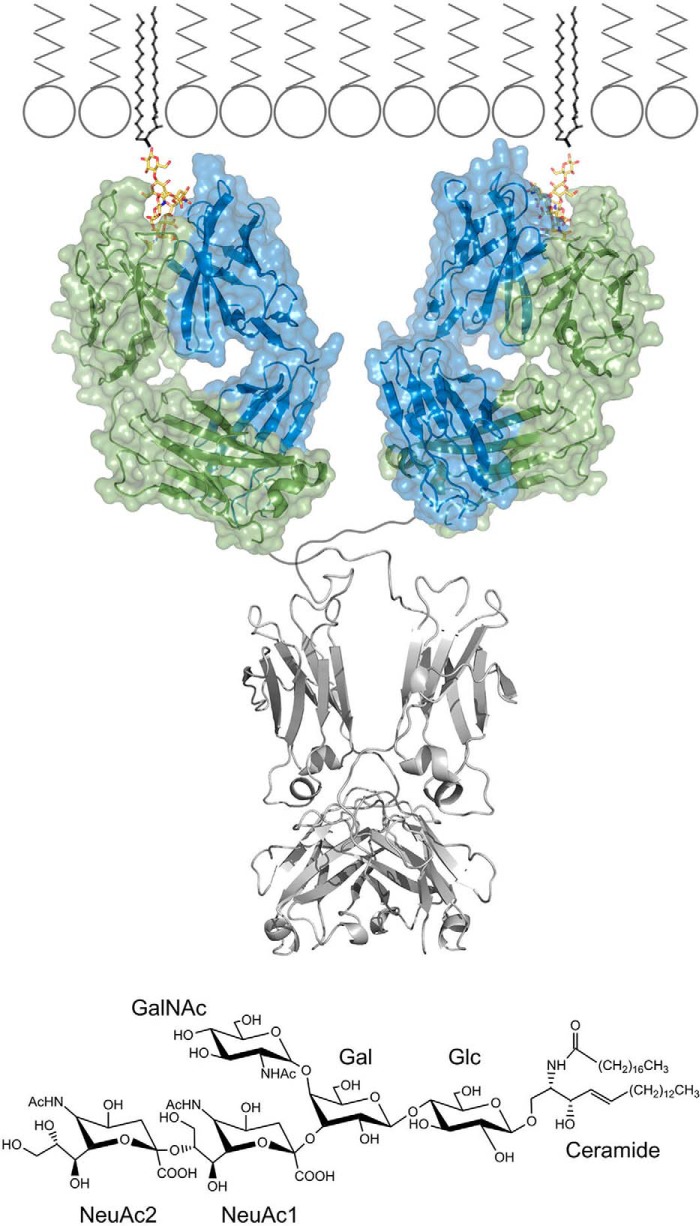
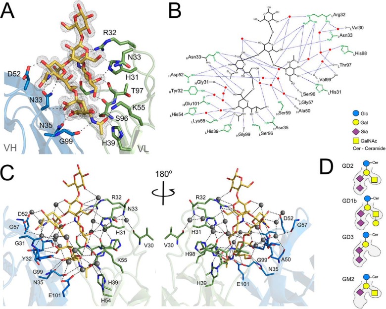
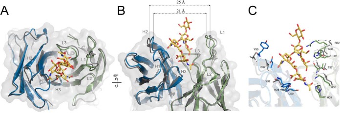
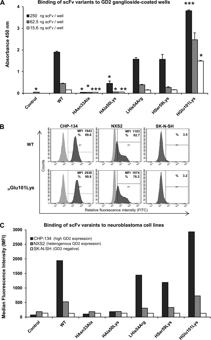
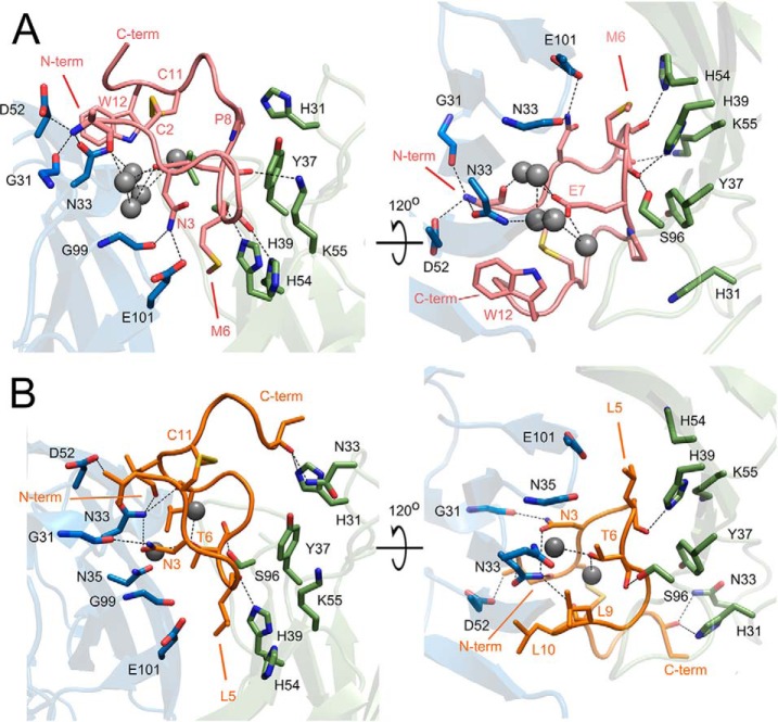
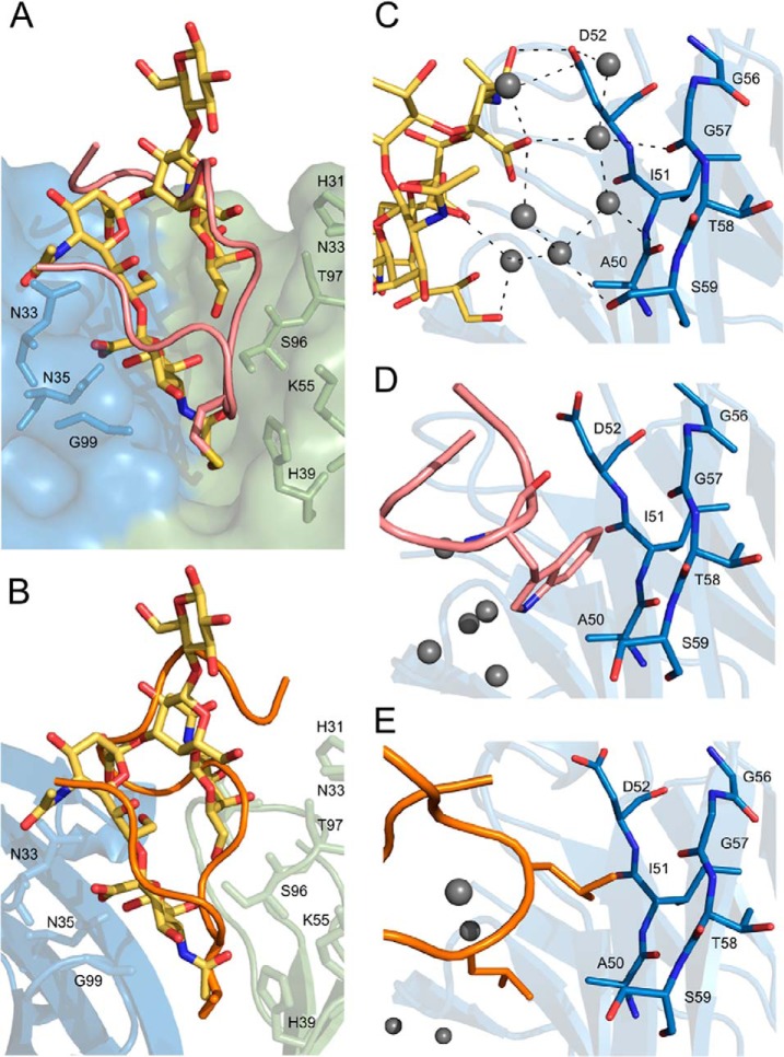
Monoclonal antibodies targeting GD2 ganglioside (GD2) have recently been approved for the treatment of high risk neuroblastoma and are extensively evaluated in clinics in other indications. This study illustrates how a therapeutic antibody distinguishes between different types of gangliosides present on normal and cancer cells and informs how synthetic peptides can imitate ganglioside in its binding to the antibody. Using high resolution crystal structures we demonstrate that the ganglioside recognition by a model antibody (14G2a) is based primarily on an extended network of direct and water molecule mediated hydrogen bonds. Comparison of the GD2-Fab structure with that of a ligand free antibody reveals an induced fit mechanism of ligand binding. These conclusions are validated by directed mutagenesis and allowed structure guided generation of antibody variant with improved affinity toward GD2. Contrary to the carbohydrate, both evaluated mimetic peptides utilize a "key and lock" interaction mechanism complementing the surface of the antibody binding groove exactly as found in the empty structure. The interaction of both peptides with the Fab relies considerably on hydrophobic contacts however, the detailed connections differ significantly between the peptides. As such, the evaluated peptide carbohydrate mimicry is defined primarily in a functional and not in structural manner.


Figures

Fig. 1.
Fig. 1.
Recognition of GD2 ganglioside by monoclonal antibody 14G2a at the cell surface. (top panel) Antigen combining region of 14G2a antibody recognizes the sugar moiety of GD2 ganglioside (yellow), which is exposed to the extracellular milieu. The lipid part of the ganglioside is buried inside the cell membrane. GD2 bound Fab structure determined in this study is shown in color. Fc fragment (PDB ID: 1igt) and membrane model derived from published data are shown in corresponding scale and colored gray. (bottom panel) Chemical structure of GD2 ganglioside and sugar ring nomenclature used throughout the study.
Fig. 2.
Fig. 2.
GD2 ganglioside binding mode at the antigen-combining site of 14G2a. (A) GD2 (yellow) recognition relies heavily on the hydrogen bond interactions with the main chain and the side chains of both the heavy (blue) and the light (green) chains of the antibody. Antibody residues contributing major interactions are shown as sticks. Hydrogen bonds are depicted as black dotted lines. Gray mesh represents Fo-Fc omit map for the ganglioside contoured at 1.5σ. (B) Schematic representation of the major interactions guiding the recognition and specificity of 14G2a antibody toward GD2 sugar. (C) Spatial view of interactions depicted schematically in panel B. (D) Schematic representation of the steric determinants that dictate the strict preference of 14G2a toward GD2, but not closely related gangliosides GD1b, GD3 and GM2.
Fig. 3.
Fig. 3.
Ganglioside binding to 14G2a follows an induced fit mechanism. (A) Overlay of ligand free (gray) and GD2 (yellow) containing (heavy and light chains in blue and green, respectively) structures of 14G2a antibody. Ganglioside binding induces narrowing of the V shaped binding groove associated with cooperative closing of antibody CDR loops around the ligand. (B) The entrance to the groove is 25 Å wide in the ligand free structure and only 21 Å wide in the ligand containing structure. The largest movement is associated with CDR loops H2 and L1, but all other loops also close around the ligand. (C) Same view as in panel B, but the sidechains that undergo most pronounced translations and rearrangements upon ligand binding are shown in ligand free (gray) and ligand bound (color) antibody structures.
Fig. 4.
Fig. 4.
Relative affinity and specificity of scFv variants toward GD2. (A) Binding of scFv variants to GD2 as evaluated by ELISA. Plastic surface was coated with GD2 and incubated with particular scFv, which binding was detected using HRP-conjugated anti-His tag antibody; WT - wild type scFv, Control - signal from wells incubated with anti-His tag antibody alone. The statistical significance in ELISA signals between the wild type scFv-14G2a and its mutants was evaluated by t test (p values are indicated as follows p < 0.05 (*), p < 0.01 (**) and p < 0.001 (***). (B, C) Binding of scFv variants to GD2 on the cell surface. CHP-134 (high GD2 expression), NXS2 (heteregenous expression of GD2) and SK-NS-H (GD2-negative) neuroblastoma cell lines were first incubated with evaluated scFv variants and then with FITC-labeled anti-His tag antibody. Staining was measured by flow cytometry. (B) Representative histograms showing binding of WT and HGlu101Lys variants of scFv (5 μg/ml) to tested neuroblastoma cells (dark gray) overlaid with control staining of the respective cells with FITC-conjugated anti-His tag antibody alone (light gray). Based on signals from cells stained with the latter antibody alone, positive cell pools were determined (gate P7) for which median fluorescence intensity (MFI) and/or percentage of staining (%) values were measured (as indicated on the plots). (C) MFI signals for single cell populations stained with particular scFv and FITC-conjugated anti-His tag antibody, or the latter antibody alone (control).
Fig. 5.
Fig. 5.
Binding of ganglioside peptide mimetics at the antigen combining site of 14G2a. Recognition of peptide 1 (A, pink) and peptide 2 (B, orange) relies on hydrogen bond and hydrophobic interactions. The peptides are represented as main chain trace and the antibody is shown in ribbon representation. For clarity, only the most important residues mediating the antibody-peptide interaction are shown in stick representation. The ligand residue located at the bottommost pocket of the binding groove is indicated by an arrow. Antibody model orientation and color coding same as in Fig. 2.
Fig. 6.
Fig. 6.
Comparison of the binding modes of GD2 and peptide mimetics at the 14G2a antigen combining site. The general disposition of the main chain of peptide 1 (A) and peptide 2 (B) imitates the overall shape of the ganglioside sugar (yellow) because of general steric constraints imposed by the antigen binding cavity. However, the detailed interactions differ significantly as exemplified: (A–B) at the bottommost pocket of the binding cavity the carbohydrate is involved primarily in hydrogen bond interactions whereas both peptides expose hydrophobic side chains. (C) The water molecule mediated interactions of GD2 with CDR H2 are replaced by hydrophobic contacts in the case of peptide 1 (D) and peptide 2 (E). (all panels) Peptides are represented as a main-chain trace with the discussed residues shown as sticks.

References

    1. Cheever M. A., Allison J. P., Ferris A. S., Finn O. J., Hastings B. M., Hecht T. T., Mellman I., Prindiville S. A., Viner J. L., Weiner L. M., Matrisian L. M. (2009) The prioritization of cancer antigens: A national cancer institute pilot project for the acceleration of translational research. Clin. Cancer Res. 15, 5323–5337 - PMC - PubMed
    1. Roth M., Linkowski M., Tarim J., Piperdi S., Sowers R., Geller D., Gill J., Gorlick R. (2014) Ganglioside GD2 as a therapeutic target for antibody-mediated therapy in patients with osteosarcoma. Cancer 120, 548–554 - PMC - PubMed
    1. Yoshida S., Fukumoto S., Kawaguchi H., Sato S., Ueda R., Furukawa K. (2001) Ganglioside G(D2) in small cell lung cancer cell lines: enhancement of cell proliferation and mediation of apoptosis. Cancer Res. 61, 4244–4252 - PubMed
    1. Wu Z. L., Schwartz E., Seeger R., Ladisch S. (1986) Expression of GD2 ganglioside by untreated primary human neuroblastomas. Cancer Res. 46, 440–443 - PubMed
    1. Cheung N. K., Dyer M. A. (2013) Neuroblastoma: developmental biology, cancer genomics and immunotherapy. Nat. Rev. Cancer 13, 397–411 - PMC - PubMed

Publication types