. 2014 Nov 24;31(4):487-502.
doi: 10.1016/j.devcel.2014.09.013. Epub 2014 Nov 6.

The chromosome axis controls meiotic events through a hierarchical assembly of HORMA domain proteins

Affiliations

The chromosome axis controls meiotic events through a hierarchical assembly of HORMA domain proteins

Yumi Kim et al. Dev Cell. .

Abstract

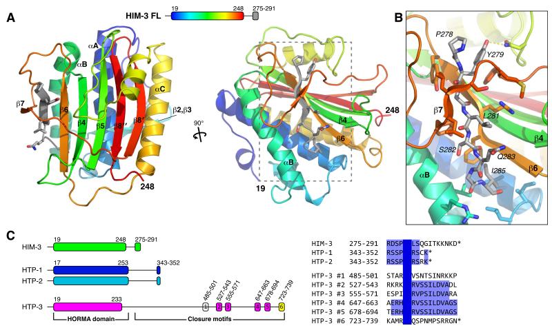
Proteins of the HORMA domain family play central, but poorly understood, roles in chromosome organization and dynamics during meiosis. In Caenorhabditis elegans, four such proteins (HIM-3, HTP-1, HTP-2, and HTP-3) have distinct but overlapping functions. Through combined biochemical, structural, and in vivo analysis, we find that these proteins form hierarchical complexes through binding of their HORMA domains to cognate peptides within their partners' C-terminal tails, analogous to the "safety belt" binding mechanism of Mad2. These interactions are critical for recruitment of HIM-3, HTP-1, and HTP-2 to chromosome axes. HTP-3, in addition to recruiting the other HORMA domain proteins to the axis, plays an independent role in sister chromatid cohesion and double-strand break formation. Finally, we find that mammalian HORMAD1 binds a motif found both at its own C terminus and at that of HORMAD2, indicating that this mode of intermolecular association is a conserved feature of meiotic chromosome structure in eukaryotes.


Figures

Figure 1
Figure 1. Structure of C. elegans HIM-3 reveals conserved “closure motifs”
(A) Two views of C. elegans HIM-3, with HORMA domain colored as a rainbow from N- to C-termini and secondary structure elements labeled according to the Mad2 convention (Luo et al., 2002; Sironi et al., 2002), with the C-terminal “closure motif” residues 278-285 in gray (see schematic, top). See Figure S1A for comparison with Mad2 and Rev7. (B) Detail view showing interactions between the closure motif and the “safety belt” of HIM-3. (C) Left: schematic of HIM-3, HTP-1, HTP-2 and HTP-3 N-terminal HORMA domains and C-terminal “closure motifs.” Right: alignment of putative closure motifs from all four C. elegans HORMA domain proteins. See also Figure S1B-D.
Figure 2
Figure 2. The C. elegans meiotic HORMA domain proteins form a complex
(A) Mass spectrometry results from HTP-3-GFP pulldowns. Pulldowns were performed in conditions that favored either soluble or chromatin-associated complexes (see Extended Experimental Procedures). (B) Size exclusion chromatography and multi-angle light scattering (SEC-MALS) analysis of HORMA domain protein complexes (a: complexes containing HTP-3 showed strong polydispersity indicative of a mixture of different-weight particles). See Figure S2 for SDS-PAGE analysis of fractions and representative SEC-MALS results. (C) SDS-PAGE analysis of co-expressed complexes purified using Strep-HTP-3 (left panel) or HIM-3-6His (right panel).
Figure 3
Figure 3. C. elegans HORMA domain proteins bind distinct closure motifs
(A-C) Fluorescence polarization (FP) peptide-binding assay for HIM-3 (A), HTP-1 (B), and HTP-2 (C). Peptides used are shown in panel (D). (D) Measured Kd’s for HIM-3, HTP-1, and HTP-2 binding different closure motifs. n.s.; no significant binding detected. (E) Schematic illustrating the closure motif binding specificities of HIM-3, HTP-1, and HTP-2.
Figure 4
Figure 4. HIM-3 binding to HTP-3 motifs #2-5 is critical for homolog synapsis and successful meiosis
(A) Structure of HIM-3 (green) bound to HTP-3 motif #4 (magenta). Molecular surface is shown in gray for the bound closure motif. (B) Detail view showing interactions between the closure motif and the “safety belt” of HIM-3. See also Figure S3. (C) Sequence alignment of HTP-3 motifs #2-#5. (D) FP peptide binding assay for HIM-3 binding HTP-3 motif #4 wild-type (as in Figure 3A) and G652K mutant. See also Figure S4. (E) Strep-tagged HTP-3, either wild-type or 4GK mutant (conserved glycine in HTP-3 motifs #2-#5 mutated to lysine; schematic at top), was coexpressed in E. coli with untagged HIM-3 (left) or HTP-1 (right), and purified using Strep-tactin resin to isolate HTP-3 and directly bound proteins. (F) Wild-type and 4GK mutant htp-3-gfp transgenes were expressed in the htp-3(tm3655)I background, and mid-pachytene nuclei stained for DNA, HTP-3:GFP, HTP-1, and HIM-3. Scale bar, 5 μm. (G) Schematic showing meiotic chromosome axis formation and synapsis. (H) Wild-type or 4GK mutant HTP-3:GFP transgenes were expressed in the htp-3(tm3655)I background, and mid-pachytene nuclei were stained for DNA, HTP-3:GFP (green), and SYP-2 (red). All images are maximum-intensity projections of deconvolved 3D image stacks. Scale bar, 5 μm. See also Figure S5.
Figure 5
Figure 5. HTP-1/HTP-2 is recruited to chromosomes by both HTP-3 and HIM-3
(A) Structure of HTP-1P84L (blue) bound to the HIM-3 closure motif (green). A molecular surface representation is shown in gray for the bound closure motif. (B-C) Detail views showing interactions between HTP-1P84L and the HIM-3 closure motif (B) or HTP-3 motif #1 (C). Leucine 84 is shown in stick view. See Figure S3 for representative electron density. (D) Structure of HTP-2 (cyan) bound to the HIM-3 closure motif (green). (E-G) Detail views showing interactions between HTP-2 and the HIM-3 closure motif (E), HTP-3 motif #1 (F), or HTP-3 motif #6 (G). (H) Sequence alignment of closure motifs specific for binding the HTP-1 and HTP-2 HORMA domains. (I) Strep-tagged HTP-3 wild-type or GK mutants (schematic at top) were coexpressed in E. coli with untagged HIM-3 (left) or HTP-1 (right), and purified using Strep-tactin resin to isolate HTP-3 and directly bound proteins. (J) Wild-type, 2GK, and 6GK mutant htp-3-gfp transgenes were expressed in the htp-3(tm3655)I background and combined with the him-3(gk149) IV null allele, and mid-pachytene nuclei stained for DNA, HTP-3:GFP, HTP-1, and HIM-3. Scale bar, 5 μm. (K) Projection images showing mid-pachytene nuclei of the strains in (J) stained for DNA, HTP-3:GFP (green), and SYP-2 (red). Scale bar, 5 μm. See also Figure S5.
Figure 6
Figure 6. Chromosome axis localization of cohesin complexes in HTP-3 closure motif GK mutants
(A) and (B) Mid-pachytene nuclei in wild-type N2, htp-3(tm3655), and htp-3(tm3655);htp-36GK worms were stained for HTP-3 and meiosis-specific kleisin subunits of the cohesin complex, REC-8 (A) or COH-3/COH-4 (B). Scale bar, 5 μm. See Figure S6 for localization of SMC subunits SMC-1 and SMC-3. (C) Projection images showing mid-pachytene nuclei stained for DNA and RAD-51 to assess the formation and repair of meiotic DSBs. (D) Schematic illustrating the major functions and proposed roles of the C. elegans meiotic HORMA domain proteins.
Figure 7
Figure 7. Mammalian HORMAD1 and HORMAD2 contain C-terminal closure motifs
(A) Schematic of human HORMAD1 and HORMAD2, with HORMA domain and C-terminal region shown as boxes. (B) Ni2+ pulldown using His6-MBP-tagged HORMAD1/2 tail segments, with untagged human HORMAD1(2-235) as prey. Top panel: 10% Load sample, Bottom panel: Ni2+-bound fraction. Red asterisks (*) indicate a contaminant in the HORMAD1 ΔC104 tail construct that is of similar molecular weight to HORMAD1(2-235). (C) Schematic of results from (B), showing that the extreme C-terminal regions of both HORMAD1 and HORMAD2 are necessary and sufficient for HORMAD1(2-235) binding. (D) Sequence alignment of putative HORMAD1 and HORMAD2 closure motifs from human (Hs) and mouse (Mm).

Comment in

References

    1. Aravind L, Koonin EV. The HORMA domain: a common structural denominator in mitotic checkpoints, chromosome synapsis and DNA repair. Trends Biochem. Sci. 1998;23:284–286. - PubMed
    1. Baumgartner B, Woltering D, Bagchi S, Larkin B, Loidl J, de los Santos T, Hollingsworth NM. Meiotic segregation, synapsis, and recombination checkpoint functions require physical interaction between the chromosomal proteins Red1p and Hop1p. Mol Cell Biol. 2000;20:6646–6658. - PMC - PubMed
    1. Blat Y, Protacio RU, Hunter N, Kleckner N. Physical and functional interactions among basic chromosome organizational features govern early steps of meiotic chiasma formation. Cell. 2002;111:791–802. - PubMed
    1. Burger J, Merlet J, Tavernier N, Richaudeau B, Arnold A, Ciosk R, Bowerman B, Pintard L. CRL2(LRR-1) E3-ligase regulates proliferation and progression through meiosis in the Caenorhabditis elegans germline. PLoS Genet. 2013;9:e1003375. - PMC - PubMed
    1. Caryl AP, Armstrong SJ, Jones GH, Franklin FC. A homologue of the yeast HOP1 gene is inactivated in the Arabidopsis meiotic mutant asy1. Chromosoma. 2000;109:62–71. - PubMed

Publication types

MeSH terms

Substances