. 2014 Dec;21(12):1091-6.
doi: 10.1038/nsmb.2905. Epub 2014 Nov 10.

Structural determinants of integrin β-subunit specificity for latent TGF-β

Affiliations

Structural determinants of integrin β-subunit specificity for latent TGF-β

Xianchi Dong et al. Nat Struct Mol Biol. 2014 Dec.

Abstract

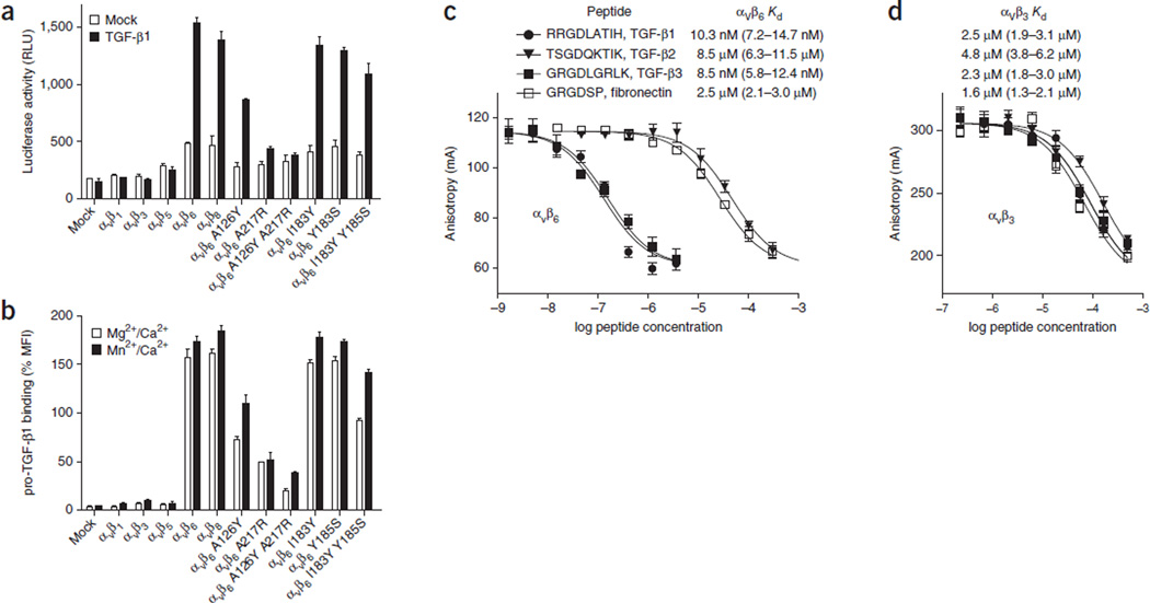
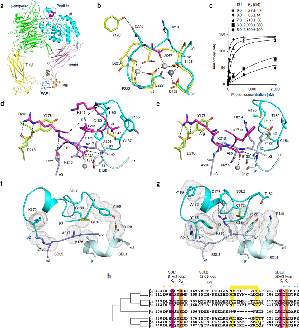
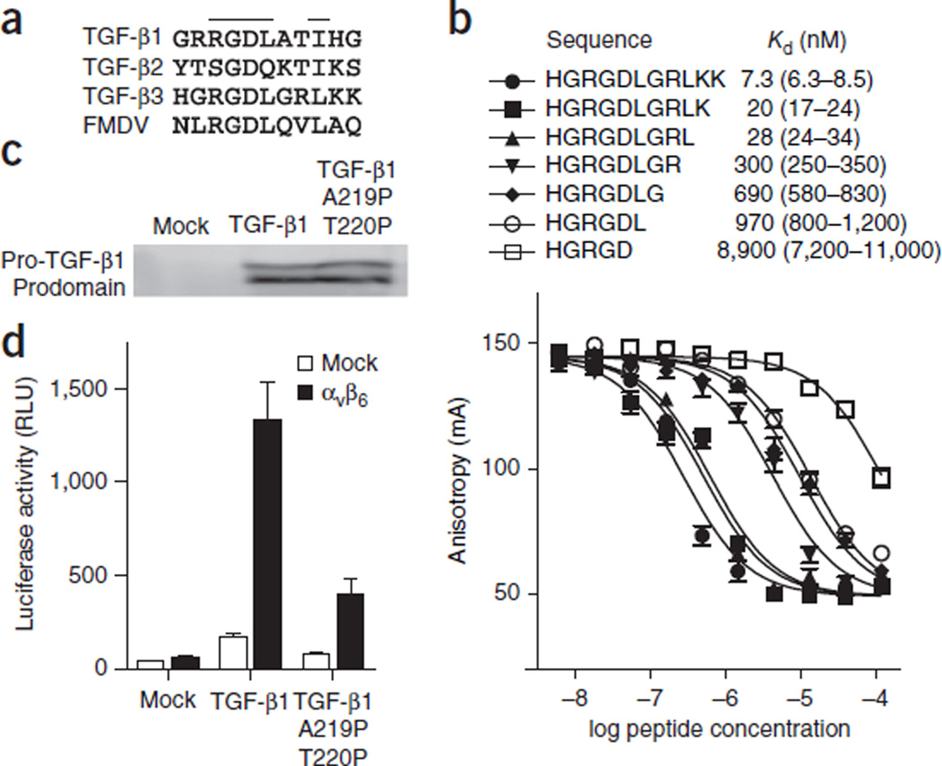
Eight integrin α-β heterodimers recognize ligands with an Arg-Gly-Asp (RGD) motif. However, the structural mechanism by which integrins differentiate among extracellular proteins with RGD motifs is not understood. Here, crystal structures, mutations and peptide-affinity measurements show that αVβ6 binds with high affinity to a RGDLXXL/I motif within the prodomains of TGF-β1 and TGF-β3. The LXXL/I motif forms an amphipathic α-helix that binds in a hydrophobic pocket in the β6 subunit. Elucidation of the basis for ligand binding specificity by the integrin β subunit reveals contributions by three different βI-domain loops, which we designate specificity-determining loops (SDLs) 1, 2 and 3. Variation in a pair of single key residues in SDL1 and SDL3 correlates with the variation of the entire β subunit in integrin evolution, thus suggesting a paradigmatic role in overall β-subunit function.


Figures

Figure 1
Figure 1
Activation and binding of pro-TGF-β1 by wild-type and mutant αV integrins. (a) Indicated HEK293T transfectants assayed for TGF-β1 activation with mink lung luciferase reporter cells, measured as relative light units (RLU). Mock, control mock transfection. (b) Saturation binding of FITC–pro-TGF-β1 to HEK293T transfectants, shown as percentage mean fluorescence intensity (% MFI) of αV P2W7 monoclonal antibody binding. Slashes denote ‘and.’ (c,d) Binding of peptides to αVβ6 (c) or αVβ3 (d) headpieces, measured by fluorescence anisotropy. Anisotropy is measured as millianisotropy units (mA), as (FF)/(F + 2F) × 1,000, where F is the fluorescence intensity parallel to the excitation plane, and F is the fluorescence intensity perpendicular to the excitation plane. Data show mean ± s.e.m. of technical triplicate samples. Peptides at the indicated concentrations were used with 200 nM αVβ6 or 4 µM αVβ3 headpiece and 5 nM of fluorescent peptide probe. Kd was calculated from IC50 as described.
Figure 2
Figure 2
Crystal structures and comparisons of the αVβ6 headpiece. (a) Overall ribbon diagram of the αVβ6 headpiece (with each domain in a different color) with pro-TGF-β3 peptide (magenta). (b) Conformational change of the βI α2-α3 loop in the absence and presence of pro-TGF-β3. Carbon color code: green, in absence of peptide αV; cyan, β6; yellow, in presence of peptide β6; magenta, peptide aspartate. Metals are white or gray spheres. (c) pH dependence of binding affinity. Binding of FITC–pro-TGF-β3 peptide measured with fluorescence anisotropy is shown. Data show mean ± s.e.m. of technical triplicate samples. (d,e) Ligand binding of αVβ6 to pro-TGF-β3 peptide (d) and αVβ3 to cilengitide (e). Carbon color code: green, αV; cyan, β3 or β6, with different shades for SDLs 1, 2 and 3; magenta, ligands. The MIDAS metal ion is a silver sphere. (f,g) Key residues that contribute to packing between SDLs 1, 2 and 3 in β6 (f) and β3 (g). SDL color code is as in d and e. Van der Waals surfaces around interacting side chains are shown as dots. (h) Phylogenetic tree for integrin β-subunit SDL sequences. Ligand-contacting residues in SDL1 and SDL3 in the X1 positions are highlighted in pink. Residues that form packing interactions of SDL1 and SDL3 with SDL2 in the X2 position are highlighted in orange. Cysteines forming disulfides are highlighted in yellow. Residues that coordinate metals are asterisked in orange (MIDAS), green (ADMIDAS) and cyan (SyMBS).
Figure 3
Figure 3
Ligands of αVβ6. (a) RGD sequences from pro-TGF-β and VP1 protein from FMDV. (b) Competitive binding affinities of TGF-β3 peptide truncations. Fluorescence anisotropy data are mean ± s.e.m. of technical triplicate samples scaled logarithmically. (c) Western blot of pro-TGF-β1 secreted by the indicated HEK293T transfectants, with antibody to the prodomain as described previously. (d) TGF-β bioassay of pro-TGF-β1 and its double-proline mutant. Data show mean ± s.e.m. of technical triplicate samples.

References

    1. Hynes RO. Integrins: bi-directional, allosteric, signaling machines. Cell. 2002;110:673–687. - PubMed
    1. Munger JS, et al. The integrin αvβ6 binds and activates latent TGFβ1: a mechanism for regulating pulmonary inflammation and fibrosis. Cell. 1999;96:319–328. - PubMed
    1. Mu D, et al. The integrin αvβ8 mediates epithelial homeostasis through MT1-MMP-dependent activation of TGF-β1. J. Cell Biol. 2002;157:493–507. - PMC - PubMed
    1. Shull MM, et al. Targeted disruption of the mouse transforming growth factor-β1 gene results in multifocal inflammatory disease. Nature. 1992;359:693–699. - PMC - PubMed
    1. Yang Z, et al. Absence of integrin-mediated TGFβ1 activation in vivo recapitulates the phenotype of TGFβ1-null mice. J. Cell Biol. 2007;176:787–793. - PMC - PubMed

Publication types

MeSH terms