. 2014 Jul 25;345(6195):459-63.
doi: 10.1126/science.1254836.

Clathrin adaptors. AP2 controls clathrin polymerization with a membrane-activated switch

Affiliations

Clathrin adaptors. AP2 controls clathrin polymerization with a membrane-activated switch

Bernard T Kelly et al. Science. .

Abstract

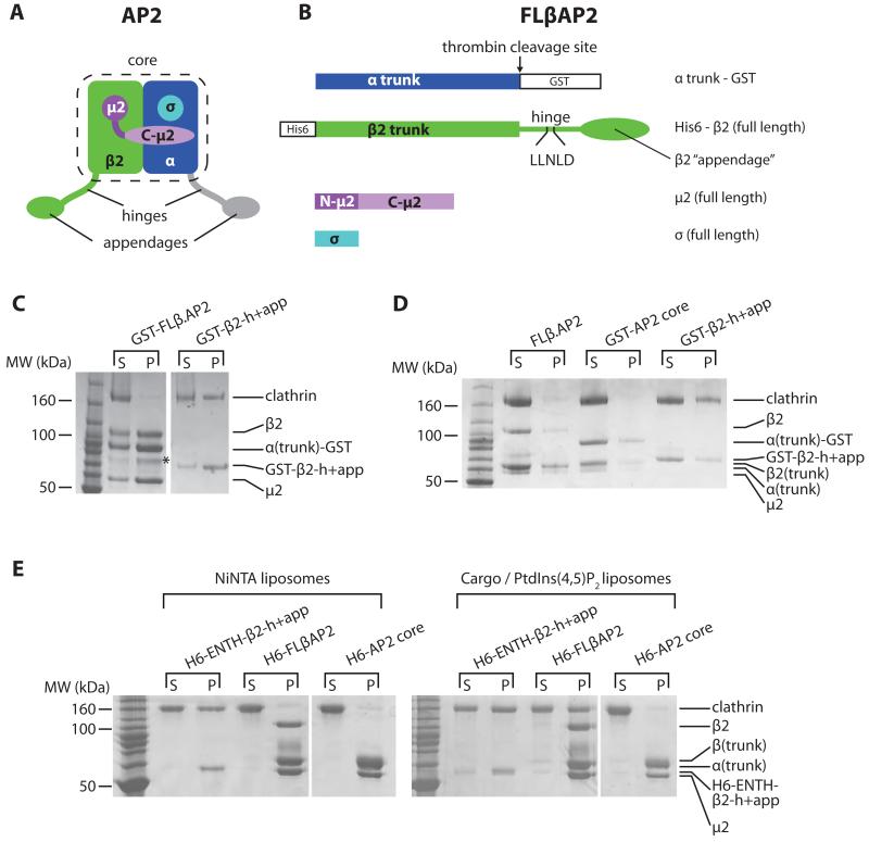
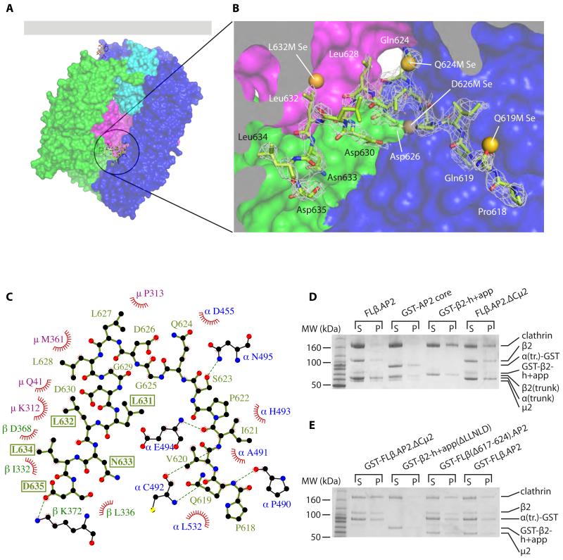
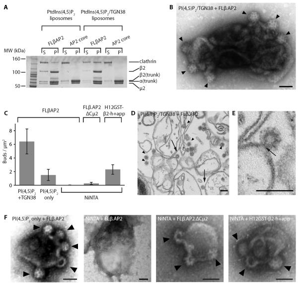
Clathrin-mediated endocytosis (CME) is vital for the internalization of most cell-surface proteins. In CME, plasma membrane-binding clathrin adaptors recruit and polymerize clathrin to form clathrin-coated pits into which cargo is sorted. Assembly polypeptide 2 (AP2) is the most abundant adaptor and is pivotal to CME. Here, we determined a structure of AP2 that includes the clathrin-binding β2 hinge and developed an AP2-dependent budding assay. Our findings suggest that an autoinhibitory mechanism prevents clathrin recruitment by cytosolic AP2. A large-scale conformational change driven by the plasma membrane phosphoinositide phosphatidylinositol 4,5-bisphosphate and cargo relieves this autoinhibition, triggering clathrin recruitment and hence clathrin-coated bud formation. This molecular switching mechanism can couple AP2's membrane recruitment to its key functions of cargo and clathrin binding.


Figures

Fig. 1
Fig. 1. AP2 can bind clathrin only when it is attached to a PtdIns(4,5)P2- and cargo-containing membrane
(A). AP2 schematic, colour-coded by subunit: α, blue; β2, green; N-μ2, dark magenta; C- μ2 pale magenta; σ, cyan. The unstructured α and β2 ‘hinge’ and ‘appendage’ subdomains, together with the ‘core’ of the complex, are indicated. Parts shown in grey are not present in FLβ.AP2. Schematics of constructs are shown in Fig S1. (B). FLβ.AP2 showing the positions of the clathrin box and purification tags. (C). Coomassie stained SDS-PAGE of glutathione-sepharose pulldowns using GST-FLβ.AP2 or GST-β2-h+app. Supernatant (‘s’) and pellet (‘p’). The band marked with an asterisk results from proteolysis of β2. (D). Coomassie stained SDS-PAGE of clathrin cage assembly assays using 2.5 μM clathrin and 1.5 μM adaptors (as indicated), overnight at 21°C, centrifuged to separate clathrin cages (‘p’) from supernatants (‘s’). (E). Coomassie stained SDS-PAGE of liposome pulldown assays. Liposomes were sequentially incubated with adaptors and clathrin, then centrifuged to separate unbound material (‘s’) from the liposome pellet (‘p’).
Fig. 2
Fig. 2. The AP2 β2 subunit LLNLD clathrin binding motif is buried in the centre of the core
(A, B). Overall (A) and closeup (B) views of the structure of βhingeHis6.AP2. The residues of the hinge resolved in the structure are shown in green as a stick representation. The AP2 core is depicted in a surface representation, coloured as in Fig. 1. The residues of the buried hinge are indicated in (B), with electron density shown as mesh (2mFo-DFc map, contoured at 0.34 e Å−3). Also shown are the positions of the selenium sites found in the bowl for each of the methionine mutants indicated, showing good agreement with the positions of the corresponding wild-type residues that were mutated. Individual LLG maps are shown in Fig S3. (C). Ligplot+ (25) diagram showing interactions between buried hinge residues (in pale green) with residues of α (blue), μ2 (magenta) and β2 (dark green). Red fans indicate hydrophobic interactions; dashed green lines indicate hydrogen bonds. The residues of the clathrin-binding motif are boxed. See also Fig. S4. (D). Clathrin cage assembly assays. (D) is identical to Fig. 1D (2.5 μM clathrin, 1.5 μM adaptors) with the addition of the FLβ.AP2.ΔCμ2 lane. (E). Assays performed as in (D) but at 28°C and with 2 μM clathrin and 4 μM adaptors as shown.
Fig. 3
Fig. 3. The ‘open’, activated form of AP2 is not compatible with the β2 hinge binding back into the core
Release of the clathrin-binding motif stimulated by conformational change. Top panels: ‘locked’AP2 core, in solution or transiently bound to the plasma membrane via PtdIns(4,5)P2 (left) and ‘open’ AP2 stably attached to the membrane via multiple PtdIns(4,5)P2s and cargo. Lower panels: views of the hinge binding site in each conformational state; in the ‘open’ state (right), the hinge residues from the ‘locked’ state βhingeH6.AP2 structure are superposed onto the ‘open’ structure and shown in grey.
Fig. 4
Fig. 4. AP2 and clathrin are sufficient to generate clathrin-coated buds on membranes
(A). Clathrin recruitment to liposomes. Synthetic liposomes supplemented with PtdIns(4,5)P2 , or with PtdIns(4,5)P2 and TGN38 peptide (as indicated), were incubated with adaptor and clathrin as shown (both at 0.4μM); supernatants (‘s’) and pellets (‘p’) were then separated and analyzed by gel electrophoresis. (B). Liposomes supplemented with PtdIns(4,5)P2 and lipid-linked TGN38 internalization incubated sequentially with FLβAP2 and clathrin, and analyzed by negative stain EM, showing clear clathrin coated membrane buds (arrowheads). (C). Number of buds formed per μm2 of membrane, estimated for various combinations of lipid and adaptor. Note that no buds were found on PtdIns(4,5)P2 liposomes ± TGN38 incubated sequentially with the AP2 core and clathrin, or with clathrin only (Fig. S9). (D, E). Liposomes supplemented with PtdIns(4,5)P2 and lipid-linked TGN38 internalization incubated sequentially with FLβAP2 and clathrin, analyzed by ultra-thin sectioning. Arrowheads indicate examples of clathrin-coated structures; arrows indicate coated buds where the connection to the liposome is clearly visible. (E) shows an enlarged image of a bud showing the “neck”. (F). Examples of liposomes supplemented with the lipids indicated (PtdIns(4,5)P2 or NiNTA-DGS) incubated sequentially with adaptors (as indicated) and clathrin, examined by negative stain EM. Note that some buds produced by FLβAP2 on PtdIns(4,5)P2-only liposomes seemed less invaginated than those shown here; this can be seen more clearly in ultrathin sections (Fig S10).

Comment in

  • Endocytosis: Unlocking AP2 activity.
    Wrighton KH. Wrighton KH. Nat Rev Mol Cell Biol. 2014 Sep;15(9):560-1. doi: 10.1038/nrm3863. Nat Rev Mol Cell Biol. 2014. PMID: 25145845 No abstract available.

References

    1. Dannhauser PN, Ungewickell EJ. Reconstitution of clathrin-coated bud and vesicle formation with minimal components. Nat. Cell Biol. 2012;14:634–639. - PubMed
    1. Cocucci E, Aguet F, Boulant S, Kirchhausen T. The first five seconds in the life of a clathrin-coated pit. Cell. 2012;150:495–507. - PMC - PubMed
    1. Traub LM. Regarding the amazing choreography of clathrin coats. PLoS Biol. 2011;9:e1001037. - PMC - PubMed
    1. Collins BM, McCoy AJ, Kent HM, Evans PR, Owen DJ. Molecular architecture and functional model of the endocytic AP2 complex. Cell. 2002;109:523–535. - PubMed
    1. Jackson LP, Kelly BT, McCoy AJ, Gaffry T, James LC, Collins BM, Höning S, Evans PR, Owen DJ. A large-scale conformational change couples membrane recruitment to cargo binding in the AP2 clathrin adaptor complex. Cell. 2010;141:1220–1229. - PMC - PubMed

Publication types

MeSH terms

Substances

Associated data