. 2016 May 6;291(19):10228-38.
doi: 10.1074/jbc.M115.700427. Epub 2016 Mar 3.

Structural and Biochemical Characterization of the Early and Late Enzymes in the Lignin β-Aryl Ether Cleavage Pathway from Sphingobium sp. SYK-6

Affiliations

Structural and Biochemical Characterization of the Early and Late Enzymes in the Lignin β-Aryl Ether Cleavage Pathway from Sphingobium sp. SYK-6

Jose Henrique Pereira et al. J Biol Chem. .

Abstract

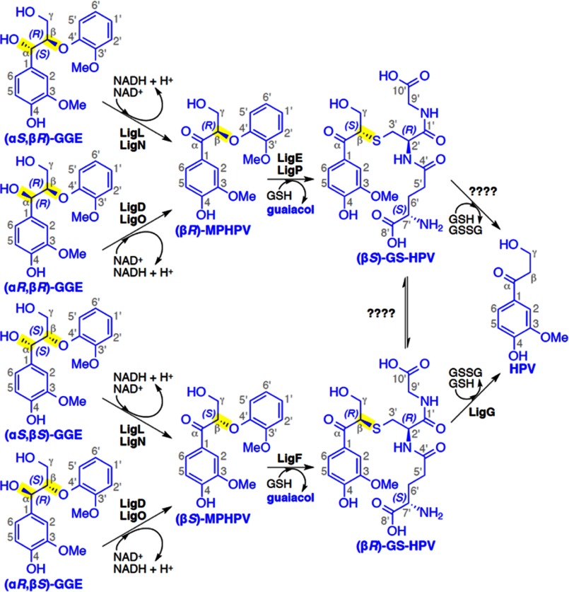
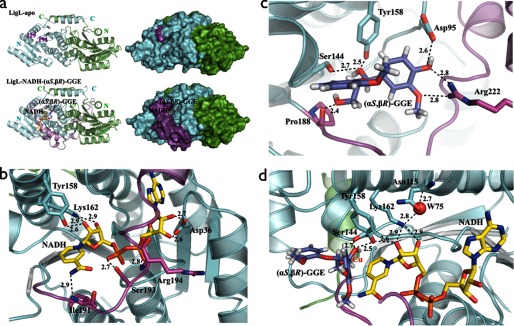
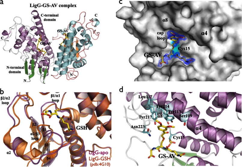
There has been great progress in the development of technology for the conversion of lignocellulosic biomass to sugars and subsequent fermentation to fuels. However, plant lignin remains an untapped source of materials for production of fuels or high value chemicals. Biological cleavage of lignin has been well characterized in fungi, in which enzymes that create free radical intermediates are used to degrade this material. In contrast, a catabolic pathway for the stereospecific cleavage of β-aryl ether units that are found in lignin has been identified in Sphingobium sp. SYK-6 bacteria. β-Aryl ether units are typically abundant in lignin, corresponding to 50-70% of all of the intermonomer linkages. Consequently, a comprehensive understanding of enzymatic β-aryl ether (β-ether) cleavage is important for future efforts to biologically process lignin and its breakdown products. The crystal structures and biochemical characterization of the NAD-dependent dehydrogenases (LigD, LigO, and LigL) and the glutathione-dependent lyase LigG provide new insights into the early and late enzymes in the β-ether degradation pathway. We present detailed information on the cofactor and substrate binding sites and on the catalytic mechanisms of these enzymes, comparing them with other known members of their respective families. Information on the Lig enzymes provides new insight into their catalysis mechanisms and can inform future strategies for using aromatic oligomers derived from plant lignin as a source of valuable aromatic compounds for biofuels and other bioproducts.

Keywords: biodegradation; biofuel; crystal structure; enzyme kinetics; lignin degradation.


Figures

FIGURE 1.
FIGURE 1.
The Sphingobium sp. strain SYK-6 β-etherase pathway. Chiral carbons at which stereospecific reactions occur are highlighted (yellow). Stereospecific reactions for (αSR)-GGE and (αSS)-GGE oxidation (by LigL and LigN), (αRR)-GGE and (αRS)-GGE oxidation (by LigD and LigO), the GSH-dependent stereospecific cleavage reactions of (βR)-MPHPV (by LigE and LigP) and (βS)-MPHPV (by LigF), and the stereospecific lyase reaction of LigG with (βR)-GS-HPV are shown.
FIGURE 2.
FIGURE 2.
Schematic representations of the biological dimers of LigD·NADH, LigO·NADH, and LigL·NADH·(αSR)-GGE, showing the overall SDR family fold composed of a central Rossmann fold. The most sequence-divergent region of SDR family members is the substrate binding loop represented in magenta. This region is disordered in all of the crystal structures solved in the apo-forms (LigD, LigO, and LigL) and in the structures of the LigD·NADH and LigO·NADH complexes. The apo-LigO and LigO·NADH structures revealed a partially ordered region with an α-helix at the N terminus of the substrate binding loop. This loop is ordered and modeled in the binary complex of LigL·NADH and ternary complex structure of LigL·NADH·(αSR)-GGE, indicating a conformational change of this loop upon cosubstrate binding.
FIGURE 3.
FIGURE 3.
a, schematic and molecular surface representations of apo-LigL and the LigL·NADH·(αSR)-GGE complex. The substrate binding loop (residues 191–229) is completely disordered in the apo-LigL structure. In the LigL·NADH·(αSR)-GGE complex structure the substrate binding loop region (magenta) acts as a lid above the NADH and GGE binding sites. b, the active site of LigL in complex with NADH showing the interactions involving the co-substrate NADH. Residue Asp36 contacts the 2′- and 3′-hydroxyl groups of the adenosine ribose sugar, the catalytic residues Tyr158 and Lys162 contact the nicotinamide ribose sugar, the nicotinamide moiety interacts with the main chain nitrogen atom from Ile191, and the phosphate groups interact with side chain atoms from Ser193 and the main chain nitrogen atom from Arg194. c, the substrate binding site for LigL·NADH·(αSR)-GGE showing residues Asp95, Ser144, Tyr158, Pro188, and Arg222 that interact directly with the GGE substrate. d, active site of LigL·NADH·(αSR)-GGE showing the catalytic tetrad N115-S144-Y158-K162 and a water molecule (W75) involved in the extended proton relay system described for the SDR family (24). Broken lines represent hydrogen bonds, and distances are shown in Å.
FIGURE 4.
FIGURE 4.
a, overall schematic representation of the LigG·GS-AV complex dimer. b, superposition of the GSH binding site of apo-LigG (magenta) and LigG-GSH (Protein Data Bank entry 4G10) (orange) structures (30). Significant conformational changes of the GSH binding site were observed on the loop regions at the N-terminal domain connecting the β1/α1, β2/α2, and α2/β3 structural elements. c, molecular surface representation of the LigG monomer in complex with the GS-AV substrate analog. A feature-enhanced map (35) contoured at 1.0 σ is shown in blue around the GS-AV substrate analog (this molecule was omitted from the model to reduce bias). The position of the catalytic Cys15 residue is highlighted in cyan. d, active site of the LigG·GS-AV complex. The glutathionyl moiety of the GS-AV substrate sits on the top of the four β-strands of the N-terminal thioredoxin domain. In addition, the AV moiety of the GS-AV molecule contacts the C-terminal α-helical domain of LigG via residues Ser109, Tyr113, and Leu117 on α4, residues Tyr214 and Tyr217 on α8, and Asn223 on the cap loop region composed of the residues 220GGGNG224.

References

    1. Sims R. E. H., Mabee W., Saddler J. N., and Taylor M. (2010) An overview of second generation biofuel technologies. Bioresour. Technol. 101, 1570–1580 - PubMed
    1. Bugg T. D. H., Ahmad M., Hardiman E. M., and Rahmanpour R. (2011) Pathways for degradation of lignin in bacteria and fungi. Nat. Prod. Rep. 28, 1883–1896 - PubMed
    1. Bugg T. D. H., Ahmad M., Hardiman E. M., and Singh R. (2011) The emerging role for bacteria in lignin degradation and bio-product formation. Curr. Opin. Biotechnol. 22, 394–400 - PubMed
    1. Masai E., Ichimura A., Sato Y., Miyauchi K., Katayama Y., and Fukuda M. (2003) Roles of the enantioselective glutathione S-transferases in cleavage of β-aryl ether. J. Bacteriol. 185, 1768–1775 - PMC - PubMed
    1. Masai E., Katayama Y., and Fukuda M. (2007) Genetic and biochemical investigations on bacterial catabolic pathways for lignin-derived aromatic compounds. Biosci. Biotechnol. Biochem. 71, 1–15 - PubMed

Publication types