. 2016 Jul 8;291(28):14639-61.
doi: 10.1074/jbc.M116.724625. Epub 2016 May 9.

Mutants of Cytochrome P450 Reductase Lacking Either Gly-141 or Gly-143 Destabilize Its FMN Semiquinone

Affiliations

Mutants of Cytochrome P450 Reductase Lacking Either Gly-141 or Gly-143 Destabilize Its FMN Semiquinone

Freeborn Rwere et al. J Biol Chem. .

Abstract

NADPH-cytochrome P450 oxidoreductase transfers electrons from NADPH to cytochromes P450 via its FAD and FMN. To understand the biochemical and structural basis of electron transfer from FMN-hydroquinone to its partners, three deletion mutants in a conserved loop near the FMN were characterized. Comparison of oxidized and reduced wild type and mutant structures reveals that the basis for the air stability of the neutral blue semiquinone is protonation of the flavin N5 and strong H-bond formation with the Gly-141 carbonyl. The ΔGly-143 protein had moderately decreased activity with cytochrome P450 and cytochrome c It formed a flexible loop, which transiently interacts with the flavin N5, resulting in the generation of both an unstable neutral blue semiquinone and hydroquinone. The ΔGly-141 and ΔG141/E142N mutants were inactive with cytochrome P450 but fully active in reducing cytochrome c In the ΔGly-141 mutants, the backbone amide of Glu/Asn-142 forms an H-bond to the N5 of the oxidized flavin, which leads to formation of an unstable red anionic semiquinone with a more negative potential than the hydroquinone. The semiquinone of ΔG141/E142N was slightly more stable than that of ΔGly-141, consistent with its crystallographically demonstrated more rigid loop. Nonetheless, both ΔGly-141 red semiquinones were less stable than those of the corresponding loop in cytochrome P450 BM3 and the neuronal NOS mutant (ΔGly-810). Our results indicate that the catalytic activity of cytochrome P450 oxidoreductase is a function of the length, sequence, and flexibility of the 140s loop and illustrate the sophisticated variety of biochemical mechanisms employed in fine-tuning its redox properties and function.

Keywords: cytochrome P45 oxidoreductase; cytochrome P450; diflavin oxidoreductase; electron transfer; flavoprotein; microsomal electron transport system; mutagenesis; oxidation-reduction (redox); redox potential.


Figures

FIGURE 1.
FIGURE 1.
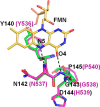
Structures of wild type CYPOR in the vicinity of the FMN isoalloxazine ring, in the oxidized state (A), reduced state (B), and a superposition of oxidized and reduced structures (C). In the oxidized structure, the five-membered 140s loop forms a double β-turn with the Gly-141–Glu-142 peptide bond adopting an O-down conformation. When the crystalline protein was reduced, the peptide bond flips to form an O-up conformation. Superposition of oxidized and reduced structures clearly shows the peptide bond isomerization (C), resulting in the formation of two H-bonds from N5 and O4 atoms of the flavin ring to the Gly-141 carbonyl oxygen and the amide nitrogen of Gly-143, respectively. These H-bonds stabilize the loop conformation, which in turn stabilizes the FMN semiquinone. In addition, when the protein is reduced, the phenolic side chain of Tyr-140 swings out from the re-face of the isoalloxazine ring, resulting in the loss of two hydrogen bonds (Tyr-140 hydroxyl to both Thr-88 and FMN phosphate oxygen). Of the two molecules in the asymmetric unit, the structure of molecule A is used for all figures, because it is better ordered than molecule B (see average B-factors in Table 2). Figs. 1–5 were generated using PyMOL Molecular Graphics System, version 1.2r3pre. Schrödinger, LLC (DeLano Scientific). Corresponding electron density map for each figure (Figs. 1–4) are included in the supplemental material.
FIGURE 2.
FIGURE 2.
Structures of ΔGly-143 CYPOR in the vicinity of the FMN ring, in the oxidized state (A) and reduced state (B), and a superposition of both structures (C). The 140s loop in this mutant is one residue shorter than that of wild type. The loop structure is extremely mobile (see supplemental Fig. S2 for electron density map). The average B-factor for the Tyr-140–Asp-144 (wild type residue numbering) loop is 69.9 Å2 (for molecule A) compared with the average B-factor of 33.6 Å2 for the entire molecule A. The disordered part of the loop is represented by thin sticks. There is no H-bond between the flavin ring and the loop. However, as in the wild type structures, there are two H-bonds from the Tyr-140 hydroxyl group to both Thr-88 and a phosphate oxygen of FMN. In the reduced structure, only the main chain atoms of Tyr-140–Gly-141of the entire 140s loop can be clearly modeled, and the rest of the loop structure shown with thin sticks represents a possible modeled structure. Furthermore, the phenol ring of Tyr-140 is completely disordered, and a possible location of the ring is shown with dotted lines (see supplemental Fig. S2 for the electron density map).
FIGURE 3.
FIGURE 3.
Structures of ΔGly-141 CYPOR in the vicinity of the FMN ring, in the oxidized state (A), reduced state (B), and a superposition of both structures (C). The 140s loop is again one residue shorter than that of wild type. This shorter loop is now closer to the FMN ring and forms a typical type I′ β-turn with the main chain carbonyl oxygen of Tyr-140 pointing down away from the FMN ring. This results in the main chain amide nitrogens of Glu-142 and Gly-143 (wild type numbering) making two H-bonds with N5 and O4 of the FMN ring, respectively. However, the γ-carboxylate of E142 adopts two conformations (shown in thin sticks), one of which is close to the FMN N5 atom (∼3.5 Å). The structure of the reduced form is essentially the same as the oxidized state, except that the Tyr-140 has two conformations. However, the γ-carboxylate of Glu-142 has now only one conformation pointing away from FMN, suggesting that the carboxylate in the alternative conformation observed in the oxidized structure would be too close to the protonated N5 of the reduced FMN.
FIGURE 4.
FIGURE 4.
Structures of ΔG141/E142N CYPOR in the vicinity of the FMN ring, in the oxidized form (A), reduced form (B), and a superposition of both structures (C). Both oxidized and reduced structures are almost identical, and they are also very similar to those of ΔGly-141, except that the Asn-142 side chain is clearly defined in the double mutant structure. As in the wild type structure, the Tyr-140 hydroxyl group makes two H-bonds with the Thr-88 and a phosphate oxygen of FMN. Furthermore, as in the ΔGly-141 structures, the two H-bonds between the flavin ring and the 140s loop (N5 and O4 of the flavin ring to the main chain amide nitrogen atoms of Asn-142 and Gly-143, respectively) are conserved in both oxidized and reduced structures.
FIGURE 5.
FIGURE 5.
Superposition of the structures of the 140s loops of ΔG141/E142N CYPOR (green carbon atoms) and P450 BM3 (purple carbons and lettering). Both structures are in the oxidized state. Hydrogen bonds between the flavin ring and the amide nitrogen atoms of the loop are shown with dashed lines. Both structures are very similar, even though the loop sequences are slightly different (YNGDPT versus YNGHPP).
FIGURE 6.
FIGURE 6.
UV-visible spectra of oxidized wild type and glycine deletion mutants of CYPOR.
FIGURE 7.
FIGURE 7.
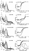
Spectral changes and a plot of absorbance at the specified wavelengths versus the potential during titration of the separate mutant FMN domains with dithionite. A, ΔGly-141. B, E142N. C, ΔG141/E142N. D, ΔGly-143. Inset shows absorbance changes at 592 nm during the titration.
FIGURE 8.
FIGURE 8.
Spectral changes of the diflavin wild type and glycine deletion mutants and the FMN domain of ΔGly-143 CYPOR during anaerobic titration with dithionite. Insets plot the change in absorbance at 454–466 nm (overall reduction) and 585–590 nm (blue semiquinone) versus the electron equivalents of dithionite added. A, wild type. B, ΔG141. Because the spectral changes at equilibrium are the same for both the ΔGly-141 and ΔG141/E142N mutants, only a single data set is illustrated. C, ΔGly-143. D, FMN domain of ΔGly-143.
FIGURE 9.
FIGURE 9.
Kinetic traces of the reduction of the diflavin wild type and three glycine deletion mutants of CYPOR by 1 molar eq of NADPH under anaerobic conditions at 25 °C. A, kinetic traces at 450–466 nm (overall reduction). B, kinetic traces at 590 nm (blue semiquinone). C, kinetic traces at 390 nm (red semiquinone). See “Experimental Procedures” for details.
FIGURE 10.
FIGURE 10.
Kinetic traces of the reduction of the diflavin wild type and three glycine deletion mutants of CYPOR by 10 molar eq of NADPH under anaerobic conditions at 25 °C. A, kinetic traces at 450–466 nm (overall reduction). B, kinetic traces at 590 nm (blue semiquinone). C, kinetic traces at 390 nm (red semiquinone). See “Experimental Procedures” for details.
SCHEME 1.
SCHEME 1.
FIGURE 11.
FIGURE 11.
Kinetics of the autoxidation of the two-electron-reduced diflavin and FMN domains of the wild type and three glycine deletion mutants at 25 °C. The reaction was monitored at the absorbance maximum of each protein as follows: wild type (452 nm); ΔGly-143 (459 nm); ΔGly-141l and ΔG141/E142N (466 nm). A, diflavin proteins; B, FMN domains. The inset compares blue semiquinone formation and decay during autoxidation of the wild type and the ΔGly-143 mutant hydroquinone at 585 nm.
FIGURE 12.
FIGURE 12.
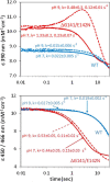
Comparison of the rate of reduction of the FMN domain of WT and ΔG141/E142N at pH 7 and 9. Absorbance changes at 390 and 450–466 nm during the reduction of the WT (blue solid line, pH 7; blue dotted line, pH 9) and mutant (red solid line, pH 7; red dotted line, pH 9) by 10 molar eq of Na2S2O4 are illustrated. The final concentration after mixing at 25 °C was 5 μm FMN domain, 50 μm Na2S2O4, 100 mm potassium phosphate buffer, 15% glycerol. Each trace is the average of three individual traces.

References

    1. Massey V., and Hemmerich P. (1980) Active-site probes of flavoproteins. Biochem. Soc. Trans. 8, 246–257 - PubMed
    1. Røhr A. K., Hersleth H.-P., and Andersson K. K. (2010) Tracking flavin conformations in protein crystal structures with Raman spectroscopy and QM/MM calculations. Angew. Chem. Int. Ed. Engl. 49, 2324–2327 - PubMed
    1. Cui D., Koder R. L. Jr., Dutton P. L., and Miller A.-F. (2011) 15N solid-state NMR as a probe of flavin H-bonding. J. Phys. Chem. B 115, 7788–7798 - PMC - PubMed
    1. Evans E. W., Dodson C. A., Maeda K., Biskup T., Wedge C. J., and Timmel C. R. (2013) Magnetic field effects in flavoproteins and related systems. Interface Focus 3, 20130037. - PMC - PubMed
    1. Miura R. (2001) Versatility and specificity in flavoenzymes: control mechanisms of flavin reactivity. Chem. Rec. 1, 183–194 - PubMed

Publication types

MeSH terms

LinkOut - more resources