. 2016 Feb 12;351(6274):725-8.
doi: 10.1126/science.aac5681.

Structural basis for histone H2B deubiquitination by the SAGA DUB module

Affiliations

Structural basis for histone H2B deubiquitination by the SAGA DUB module

Michael T Morgan et al. Science. .

Abstract

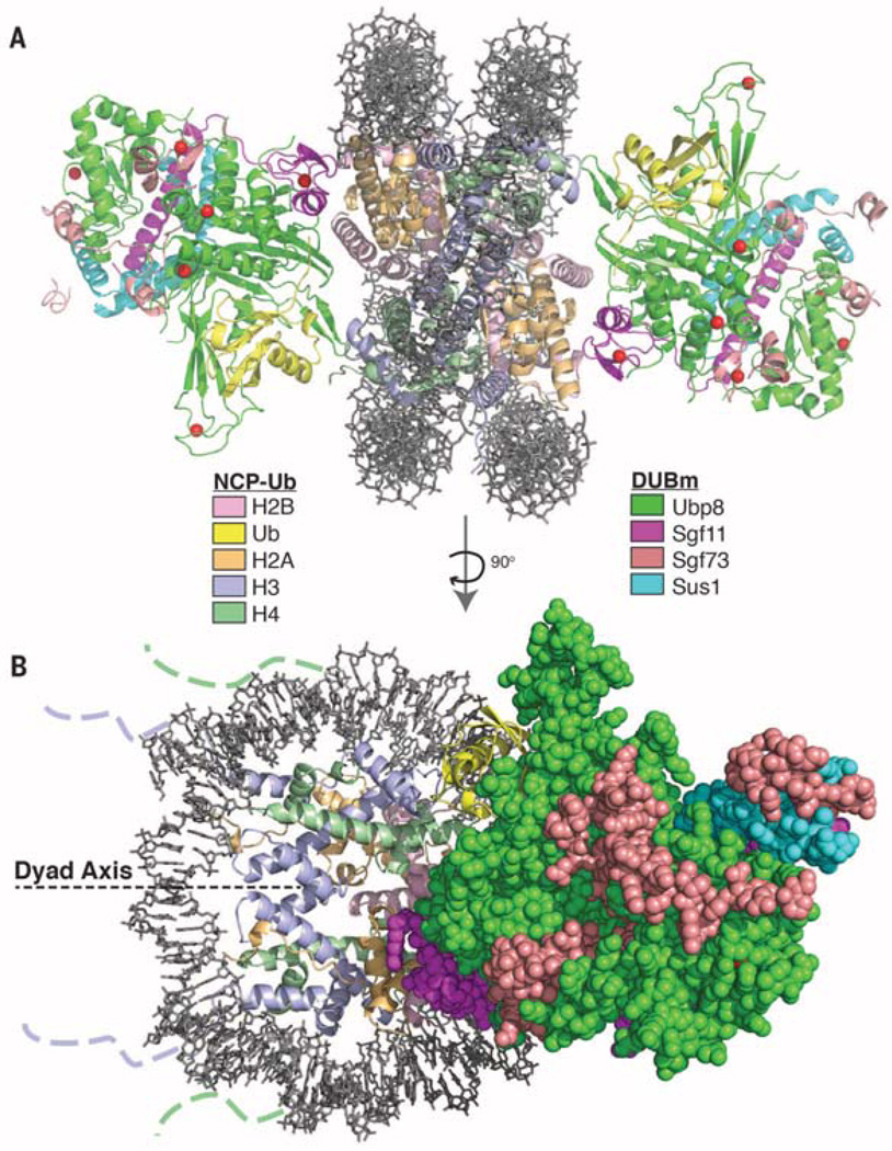
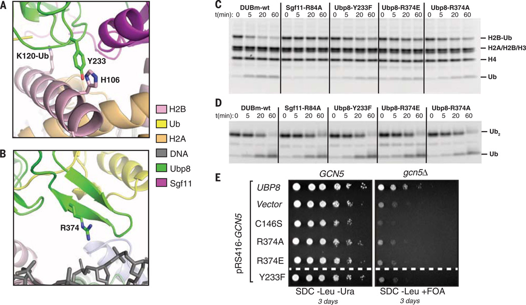
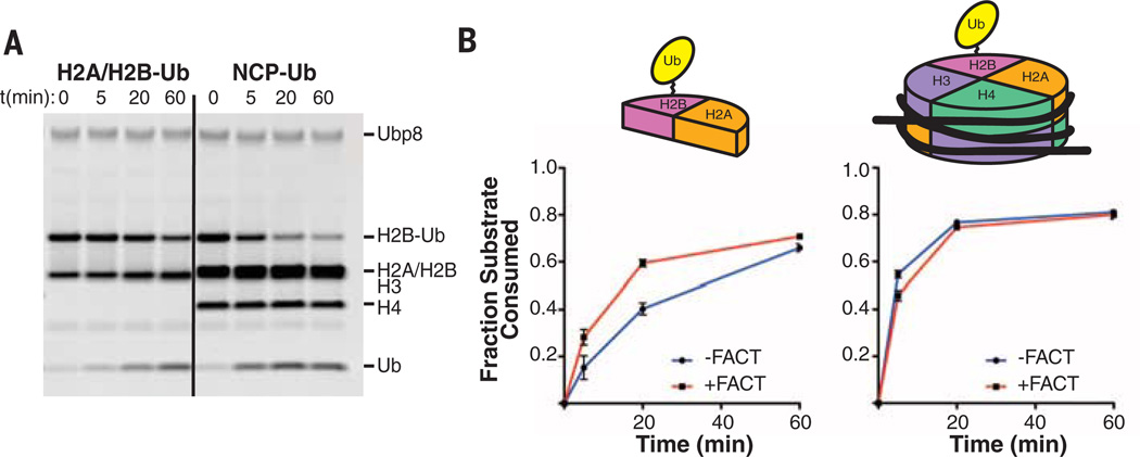
Monoubiquitinated histone H2B plays multiple roles in transcription activation. H2B is deubiquitinated by the Spt-Ada-Gcn5 acetyltransferase (SAGA) coactivator, which contains a four-protein subcomplex known as the deubiquitinating (DUB) module. The crystal structure of the Ubp8/Sgf11/Sus1/Sgf73 DUB module bound to a ubiquitinated nucleosome reveals that the DUB module primarily contacts H2A/H2B, with an arginine cluster on the Sgf11 zinc finger domain docking on the conserved H2A/H2B acidic patch. The Ubp8 catalytic domain mediates additional contacts with H2B, as well as with the conjugated ubiquitin. We find that the DUB module deubiquitinates H2B both in the context of the nucleosome and in H2A/H2B dimers complexed with the histone chaperone, FACT, suggesting that SAGA could target H2B at multiple stages of nucleosome disassembly and reassembly during transcription.


Figures

Fig. 1
Fig. 1. Overview of the SAGA DUB module bound to the monoubiquitinated nucleosome core particle (NCP-Ub)
(A) Two DUB modules bind to each NCP-Ub. The catalytic Ubp8 USP domain and the Sgf11 zinc finger contact the substrate. (B) View of the complex perpendicular to (A). The nucleosome dyad axis is indicated. Dotted lines indicate where the N-terminal tails of histone H3 and H4 protrude from the DNA gyres.
Fig. 2
Fig. 2. The DUB module binds the acidic patch of the H2A/H2B heterodimer using the Sgf11 ZnF
(A) View of interface between the DUB module and nucleosome showing the electrostatic surface potential of the histone octamer (red, negative; blue, positive). Dashed box indicates region shown in (B). (B) Modeled contacts between Sgf11 residues R78, R84, and R91 and acidic patch residues H2A-E64, H2B-E107, and H2A-E61. The ubiquitinated lysine, H2B-K120, is indicated. (C) Effect of Sgf11 ZnF mutations on deubiquitination of nucleosomal H2B (NCP-Ub). Monoubiquitinated nucleosomes (2 µM) were incubated with 200 nM wild-type or mutant DUB module for the indicated time. (D) Effect of DUB module mutations shown in (C) on cleavage of 2 µM K48-linked diubiquitin (K48-Ub2). (E) Effect of acidic patch mutation, H2A-E64R, on deubiquitination of nucleosomal H2B by the DUB module. Conditions as in (C).
Fig. 3
Fig. 3. Contribution of Ubp8 residues to DUB module activity on nucleosomes
(A) Ubp8-Y233, which is in a position to form a potential interaction with H2B-H106. (B) Modeling showing a potential contact between Ubp8 residue R374 and nucleosomal DNA. (C) Activity assay as in Fig. 2C showing effect of Ubp8 mutations Y233F, R374E, and R374A on DUB module activity on H2B-Ub in nucleosomes. Mutant Sgf11-R84A is shown for comparison. (D) Activity of DUB module mutants shown in (C) on cleavage of 2 µM K48-linked diubiquitin (Ub2). (E) Synthetic growth assay for effect of Ubp8 mutations in combination with deletion of Gcn5.
Fig. 4
Fig. 4. Activity of the DUB module on monoubiquitinated nucleosomes versus H2A/H2B-Ub in the presence and absence of FACT
(A) Time course showing cleavage of H2B-Ub by the DUB module (200 nM) in H2A/H2B dimers (H2A/H2B-Ub, 2 µM) or nucleosomes containing H2B-Ub (NCP-Ub, 1 µM). (B) Time course comparing deubiquitination of H2A/H2B-Ub (left) and NCP-Ub (right) in the presence and absence of the FACT complex. Reactions were performed in triplicate; fraction of H2B-Ub consumed was determined by quantitating bands on gels shown in fig. S9).

Comment in

References

    1. Bonnet J, Devys D, Tora L. Drug Discov. Today. Technol. 2014;12:e19–e27. - PubMed
    1. Fuchs G, Oren M. Biochim. Biophys. Acta. 2014;1839:694–701. - PubMed
    1. Wood A, et al. Mol. Cell. 2003;11:267–274. - PubMed
    1. Sun ZW, Allis CD. Nature. 2002;418:104–108. - PubMed
    1. McGinty RK, Kim J, Chatterjee C, Roeder RG, Muir TW. Nature. 2008;453:812–816. - PMC - PubMed

Publication types

MeSH terms

Associated data