. 2015 Oct;192(1):88-99.
doi: 10.1016/j.jsb.2015.08.009. Epub 2015 Aug 18.

Polyhedra structures and the evolution of the insect viruses

Affiliations

Polyhedra structures and the evolution of the insect viruses

Xiaoyun Ji et al. J Struct Biol. 2015 Oct.

Abstract

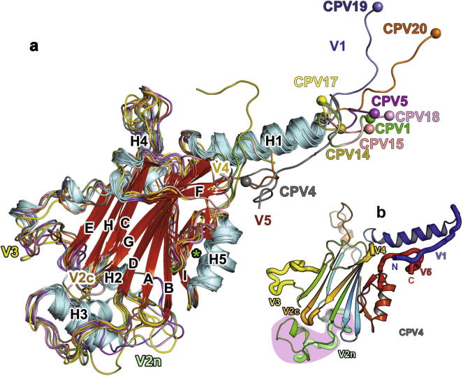
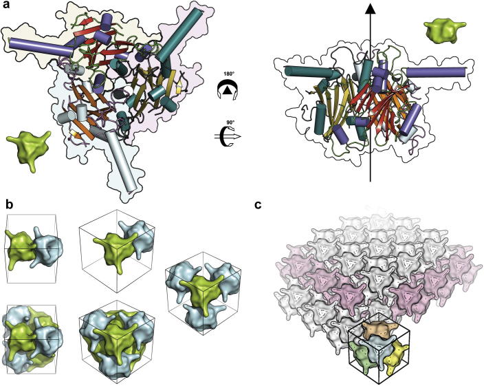
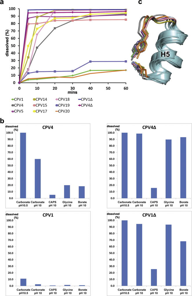
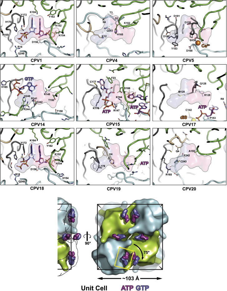
Polyhedra represent an ancient system used by a number of insect viruses to protect virions during long periods of environmental exposure. We present high resolution crystal structures of polyhedra for seven previously uncharacterised types of cypoviruses, four using ab initio selenomethionine phasing (two of these required over 100 selenomethionine crystals each). Approximately 80% of residues are structurally equivalent between all polyhedrins (pairwise rmsd ⩽ 1.5 Å), whilst pairwise sequence identities, based on structural alignment, are as little as 12%. These structures illustrate the effect of 400 million years of evolution on a system where the crystal lattice is the functionally conserved feature in the face of massive sequence variability. The conservation of crystal contacts is maintained across most of the molecular surface, except for a dispensable virus recognition domain. By spreading the contacts over so much of the protein surface the lattice remains robust in the face of many individual changes. Overall these unusual structural constraints seem to have skewed the molecule's evolution so that surface residues are almost as conserved as the internal residues.

Keywords: Cypovirus; In vivo crystals; Micro focus crystallography; Polyhedra; Protein microcrystals.


Figures

Fig. 1
Fig. 1
Overall structure. (a) Cartoon representation of the structures of the polyhedrin subunit for 9 cypovirus types. Structures were aligned using SHP (Stuart et al., 1979). Secondary structure elements are marked and coloured cyan for helices and red for β-sheet with the loops coloured by cypovirus type; CPV1, green; CPV4, grey; CPV5, magenta; CPV14, olive; CPV15, salmon; CPV17 yellow; CPV18, pink; CPV19, slate; CPV20, orange. N-termini are highlighted by a sphere. The variable regions are labelled V1–V5. (b) Cartoon representation of the CPV4 polyhedrin subunit coloured from blue to red, N-terminus to C-terminus. Variable regions are highlighted by thickened “worms” and are labelled. The green star by H5 marks the conserved interaction shown in Fig. 3c.
Fig. 2
Fig. 2
From trimer to crystal. (a) Two views of a cypovirus trimer coloured by secondary structure. In the left panel (viewing the trimer from the ‘top’) each polyhedrin monomer is delineated by a different background colour. The arrow on the right panel goes through the threefold axis. The smaller green trimers are for orientation aids for panel (b). (b) Trimers can come together progressively. Trimers facing ‘inwards’ and ‘outwards’ have been coloured cyan and green respectively to aid interpretation and show the view. The unit cell is marked for clarity although it is unlikely that assembly will occur solely within it. (c) Multiple copies of the dodecamer are arrayed in the crystal. For one dodecamer each trimer is coloured individually. For clarity the dodecamers at the 1/2, 1/2, 1/2 position are coloured light pink. For all the panels the ‘core’ structure shown is that of CPV5 using just the residues which are aligned between all the 9 polyhedrin structures. Structural alignments were performed with SHP (Stuart et al., 1979). The surface maps have been progressively smoothed which results in a slightly coarse and stylised rendering but which helps to visualise the complicated interactions.
Fig. 3
Fig. 3
Polyhedra dissolution. (a) Time course of alkaline disruption of cypovirus polyhedra. The polyhedra of nine cypovirus types plus two deletion mutants were suspended in 50 mM carbonate buffer, pH 10.5 at room temperature and the non-dissolved material monitored by absorbance at 600 nm. This was converted to % disruption by comparison to the starting value. (b) Effect of five alkali pH buffers on polyhedra of 4 cypovirus types. Release of protein from insoluble polyhedra was monitored by A280 after 20 min. As a reference, aliquots of polyhedra were completely dissolved in carbonate buffer pH 12.0, and this was used to convert to % disruption. (c) Tyr-glu interaction conserved in 8 CPV types. The green star in Fig. 1a helps locate this panel to the overall structure (the views are identical). Colours of CPV types as in Fig. 1a.
Fig. 4
Fig. 4
Purine binding. The bottom panel helps orient the reader to the position of the depicted in the other panels. Side and face-on views of a unit cell are shown, with smoothed maps representing polyhedrin trimers similar to Fig. 2. In the side view the 2 trimers closest to the viewer are shown only as outlines so that the close proximity of the NTPs to the surface of the unit cell can be clearly seen. To improve clarity the positions of the ATP (magenta) and GTP (blue) moieties for just CPV1 are shown in this panel. As a reference the positions of the CPV1 ATP and GTP have been replicated across the other panels. The yellow box shows the area represented in other 9 panels. For the nine cypovirus types the same viewpoint is shown. Protein chains are coloured separately and consistently between types. Side chains of residues which either interact with nucleotides or occupy nucleotide pockets are shown. The view has narrow clipping planes to facilitate interpretation. The positions of the calcium ion in CPV5 and C142 in CPV17 are indicated.
Fig. 5
Fig. 5
Pyrimidine binding. The lower panel orientates the reader to the positions of the CTPs and UTPs within the unit cell which cluster around the centre and the apexes. The yellow box shows the area represented in other 9 panels. The view for the nine cypovirus types is from the centre out along a body diagonal. Each trimer is roughly denoted by lines. The 9 subunit protein chains visible in the view are coloured separately with the shading grouped according to which trimer they belong to (blue, green and red-yellow). Nucleotide moieties are shown as sticks along with side chains important in this region. The 13 residues missing from the CPV15 model are shown by dashed lines.
Fig. 6
Fig. 6
Phylogenetic tree showing relationship between the polyhedrin proteins. Superpositions were performed using SHP (Stuart et al., 1979) and the phylogenetic tree calculated with PHYLIP (Felsenstein, 1989). The length of the branch to baculovirus polyhedrin (BV) has been reduced to 20% of its actual length. Structures of the proteins are shown coloured from N-terminus to C-terminus, blue to red. NTPs are represented in thickened stick style with a black outline to aid visualization. Cysteines involved in disulphide bridges are shown as black spheres, and the calcium ion in CPV5 as a pink sphere.

References

    1. Abrahams J.P., Leslie A.G. Methods used in the structure determination of bovine mitochondrial F1 ATPase. Acta Crystallogr. D Biol. Crystallogr. 1996;52:30–42. - PubMed
    1. Adams P.D., Afonine P.V., Bunkoczi G., Chen V.B., Davis I.W., Echols N., Headd J.J., Hung L.-W., Kapral G.J., Grosse-Kunstleve R.W., McCoy A.J., Moriarty N.W., Oeffner R., Read R.J., Richardson D.C., Richardson J.S., Terwilliger T.C., Zwart P.H. PHENIX: a comprehensive Python-based system for macromolecular structure solution. Acta Crystallogr. D. 2010;66:213–221. - PMC - PubMed
    1. Anduleit K., Sutton G., Diprose J.M., Mertens P.P., Grimes J.M., Stuart D.I. Crystal lattice as biological phenotype for insect viruses. Protein Sci. 2005;14:2741–2743. - PMC - PubMed
    1. Attoui, H., Mertens, P.P.C., Becnel, J., Belaganahalli, S., Bergoin, M., Brussaard, C.P., Chappell, J.D., Ciarlet, M., del Vas, M., Dermody, T.S., Dormitzer, P.R., Duncan, R., Fang, Q., Graham, R., Guglielmi, K.M., Harding, R.M., Hillman, B., Makkay, A., Marzachi, A.C., Matthijnssens, J., Milne, R.G., Mohd Jaafar, F., Mori, H., Noordeloos, A.A., Omura, T., Patton, J.T., Rao, S., Maan, M., Stoltz, D., Suzuki, N., Upadhyaya, N.M., Wei, C., Zhou, H., 2012. Cypovirus, p. 572–580, in: A. M. Q. King, et al., Eds.), Virus Taxonomy: classification and nomenclature of viruses. Ninth Report of the International Committee on Taxonomy of Viruses, p, Academic Press, London.
    1. Axford D., Ji X., Stuart D.I., Sutton G. In cellulo structure determination of a novel cypovirus polyhedrin. Acta Crystallogr. D Biol. Crystallogr. 2014;70:1435–1441. - PMC - PubMed

Publication types

MeSH terms

Substances

LinkOut - more resources