. 2016 Jun 10;291(24):12641-12657.
doi: 10.1074/jbc.M116.721050. Epub 2016 Apr 1.

i-bodies, Human Single Domain Antibodies That Antagonize Chemokine Receptor CXCR4

Affiliations

i-bodies, Human Single Domain Antibodies That Antagonize Chemokine Receptor CXCR4

Katherine Griffiths et al. J Biol Chem. .

Abstract

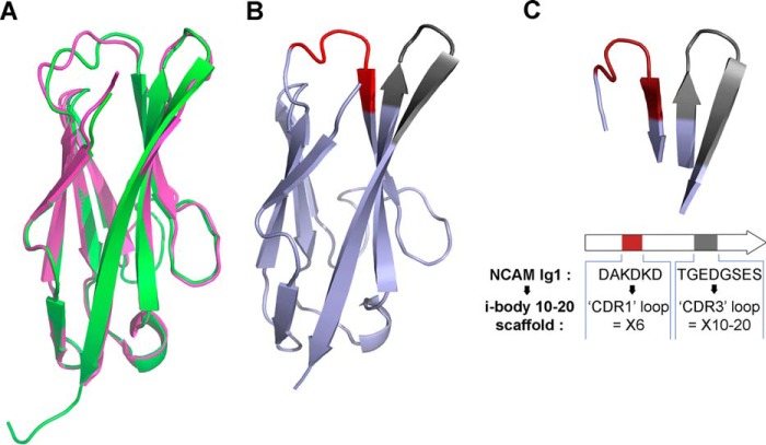
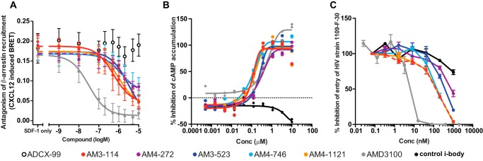
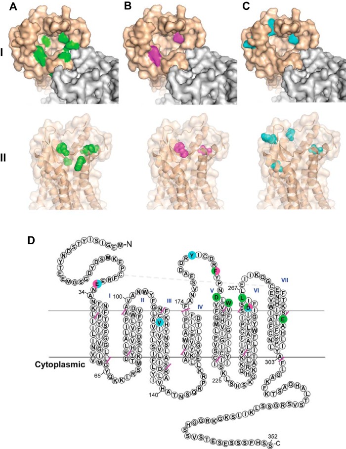
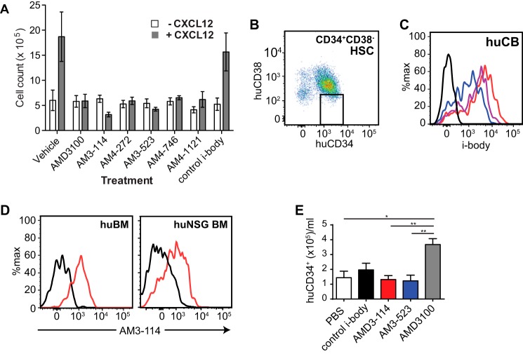
CXCR4 is a G protein-coupled receptor with excellent potential as a therapeutic target for a range of clinical conditions, including stem cell mobilization, cancer prognosis and treatment, fibrosis therapy, and HIV infection. We report here the development of a fully human single-domain antibody-like scaffold termed an "i-body," the engineering of which produces an i-body library possessing a long complementarity determining region binding loop, and the isolation and characterization of a panel of i-bodies with activity against human CXCR4. The CXCR4-specific i-bodies show antagonistic activity in a range of in vitro and in vivo assays, including inhibition of HIV infection, cell migration, and leukocyte recruitment but, importantly, not the mobilization of hematopoietic stem cells. Epitope mapping of the three CXCR4 i-bodies AM3-114, AM4-272, and AM3-523 revealed binding deep in the binding pocket of the receptor.

Keywords: CXC chemokine receptor type 4 (CXCR-4); G protein-coupled receptor (GPCR); antibody engineering; hematopoietic stem cells; protein structure; single-domain antibody (sdAb, nanobody).


Figures

FIGURE 1.
FIGURE 1.
Attributes of the i-body scaffold. A, overlay of the crystal structure of 21H-5 (human NCAM1, Ig1 domain (clone 21H-5, green)) and rat NCAM1 Ig1 (PDB 1QZ1, pink). B, ribbon diagram of the crystal structure of 21H-5 (human NCAM1, Ig1 domain) showing in red and gray the loops designated as CDR1 and CDR3, respectively. C, red and gray loops extending from the top of the molecule were replaced, respectively, with fully randomized 6-residue “CDR1” and 10–20-residue “CDR3” antigen-binding loops to produce the i-body scaffold.
FIGURE 2.
FIGURE 2.
Isolation of CXCR4 binders. A, ELISA of phage particles from sequential panning rounds, showing enrichment of i-bodies with specificity for CXCR4 lipoparticles. B, kinetic data set collected for AM3-114 binding to immobilized CXCR4. Injected concentrations were 81, 27, 9, 3, and 1 nm. Binding responses (black sensorgrams) are overlaid with fits of a simple 1:1 kinetic interaction model (orange lines). C, sequence alignment of CXCR4 i-bodies. ADCX-99 was isolated from the primary i-body library. Clones from the first affinity-matured library were AM1-126 and AM1-320. Clones from the second affinity-matured library were AM3-114, AM5-245, AM4-272, AM3-466, AM3-523, AM4-613, AM4-661, AM4-746, AM3-920, and AM4-1121. The sequence LOGO motifs show sequence conservation of residues within the CDRs.
FIGURE 3.
FIGURE 3.
Titration of AM3-114 binding to CXCR4-positive cancer cell lines. A, percent of various cell lines stained with 12G5, indicating relative expression level of CXCR4 for each cell line. B, Namalwa cells were treated with anti-CXCR4 mAb 12G5 or AM3-114 at various concentrations, and then staining was analyzed by flow cytometry. C, various cell lines were treated with AM3-114 at 10 μm, orange trace; 1 μm, blue trace; 0.001 μm, red trace, and then staining was visualized by flow cytometry.
FIGURE 4.
FIGURE 4.
Chemokine receptor screen with CXCR4 i-bodies. β-Arrestin activation following chemokine receptor stimulation in the presence of ADCX-99, AM1-126, AM1-320, AM3-114, AM4-272, AM3-523, AM4-746, AM4-1121, and AMD3100. The assay was conducted once with i-bodies, and AMD3100 was tested at a single concentration.
FIGURE 5.
FIGURE 5.
Characterization of CXCR4 i-bodies in β-arrestin, cAMP, and HIV assays. A, inhibition of β-arrestin recruitment to CXCR4 in HEK293FT cells transiently transfected with CXCR4/Rluc8 and β-arrestin2/Venus as measured by BRET. Cells were stimulated for ∼20 min with 100 nm CXCL12 in the presence of increasing concentrations of ADCX-99, AM3-114, AM4-272, AM3-523, AM4-746, AM4-1121, and AMD3100. Error bars show S.E. B, % inhibition of cAMP production in CHO-K1 cells expressing CXCR4 following stimulation with CXCL12 at EC80 (3.2 nm), in the presence of AM3-114, AM4-272, AM3-523, AM4-746, AM4-1121, and AMD3100. The line of best fit connects the duplicate data points for each test condition. C, % inhibition of entry of HIV luciferase reporter viruses pseudotyped with 1109-F-30 Env in the presence of AM3-114, AM4-272, AM3-523, AM4-746, AM4-1121, and AMD3100 and a control i-body. Error bars show S.D.
FIGURE 6.
FIGURE 6.
Epitope mapping i-body binding to CXCR4. Epitope mapping of i-bodies AM3-114, AM4-272, and AM3-523 binding to CXCR4 is shown. The primary, secondary, and tertiary contact residues identified for binding of AM3-114 (A), AM4-272 (B), and AM3-523 (C) are highlighted on space-filled depictions of the top view (panel I) and side view (panel II) of the active site of CXCR4 (derived from PDB 3ODU). D, snake plot representation of CXCR4 (derived from PDB 3ODU) showing residues involved in binding to AM3-114 (green), AM4-272 (magenta), and AM3-523 (cyan). The epitope of AM3-114 consists of residues in ECLs 1–3, as well as transmembrane (TM) regions 4 and 7. The epitope of AM4-272 consists of residues in the N terminus, ECL2, ECL3, and TM4. The epitope of AM3-523 consists of residues in N terminus, ECL2, ECL3, and TM3. Pink dashes represent membrane boundaries.
FIGURE 7.
FIGURE 7.
CXCR4 i-bodies bind but do not mobilize human stem and progenitor cells. A, in a murine model, CXCR4 i-bodies blocked cell migration into an artificial skin air-pouch, n = 5; error bars are expressed as S.E. B, representative dot plot of human CD34+CD38 HSC. C, representative flow cytometric histogram of i-body binding to human CB CD34+CD38 HSC using AM3-114 (red), AM4-272 (purple), and AM3-523 (blue). D, representative histogram of AM3-114 (red) binding to human BM CD34+CD38 HSC and muCD45huCD45+CD34+CD38 HSC from huNSG BM. Control i-body shown in black. E, mobilization of muCD45huCD45+CD34+ stem and progenitor cells in huNSG mice. Error bars are expressed as S.E. *, p < 0.05; **, p < 0.01, n = 3.
FIGURE 8.
FIGURE 8.
CXCR4 i-bodies do not mobilize murine stem and progenitor cells. A, representative flow cytometry plot of BM LSK progenitors. B, representative histogram of i-body binding to sorted murine BM LSK cells using AM3-114 (red), AM4-272 (purple), and AM3-523 (blue). C, data in B expressed as fold-increase mean fluorescence intensity (MFI) relative to control i-body. D, representative dot plot of mobilized PB LSK cells. E, total WBC. F, LSK cell content in PB of mice administered PBS, i-bodies, or AMD3100. Error bars expressed as S.E. *, p < 0.05; ****, p < 0.001, n = 3.

References

    1. Shukra A. M., Sridevi N. V., Dev C., and Kapil M. (2014) Production of recombinant antibodies using bacteriophages. Eur. J. Microbiol. Immunol. 4, 91–98 - PMC - PubMed
    1. Jost C., and Plückthun A. (2014) Engineered proteins with desired specificity: DARPins, other alternative scaffolds and bispecific IgGs. Curr. Opin. Struct. Biol. 27, 102–112 - PubMed
    1. Weidle U. H., Auer J., Brinkmann U., Georges G., and Tiefenthaler G. (2013) The emerging role of new protein scaffold-based agents for treatment of cancer. Cancer Genomics Proteomics 10, 155–168 - PubMed
    1. Wurch T., Pierré A., and Depil S. (2012) Novel protein scaffolds as emerging therapeutic proteins: from discovery to clinical proof-of-concept. Trends Biotechnol. 30, 575–582 - PubMed
    1. Rasmussen S. G., Choi H. J., Fung J. J., Pardon E., Casarosa P., Chae P. S., Devree B. T., Rosenbaum D. M., Thian F. S., Kobilka T. S., Schnapp A., Konetzki I., Sunahara R. K., Gellman S. H., Pautsch A., et al. (2011) Structure of a nanobody-stabilized active state of the β(2) adrenoceptor. Nature 469, 175–180 - PMC - PubMed

Publication types

MeSH terms