. 2016 Jun 3;291(23):12223-32.
doi: 10.1074/jbc.M116.719815. Epub 2016 Apr 18.

X-ray Crystallographic Structure of Thermophilic Rhodopsin: IMPLICATIONS FOR HIGH THERMAL STABILITY AND OPTOGENETIC FUNCTION

Affiliations

X-ray Crystallographic Structure of Thermophilic Rhodopsin: IMPLICATIONS FOR HIGH THERMAL STABILITY AND OPTOGENETIC FUNCTION

Takashi Tsukamoto et al. J Biol Chem. .

Abstract

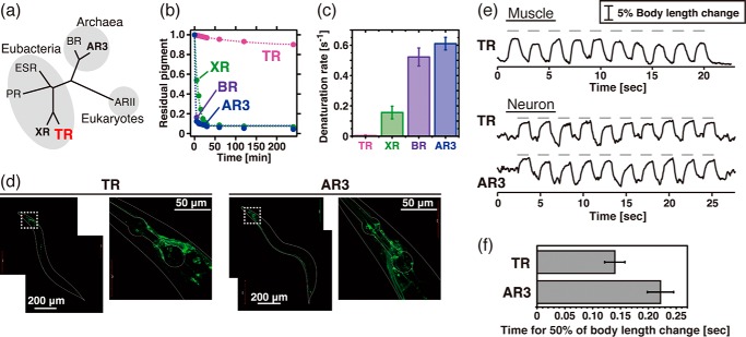
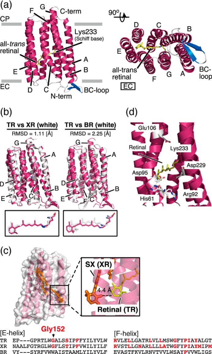
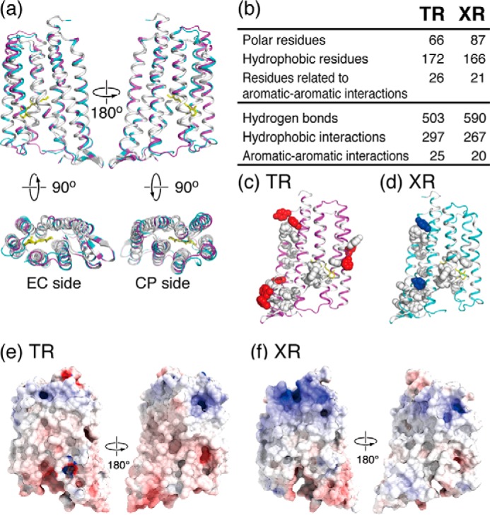
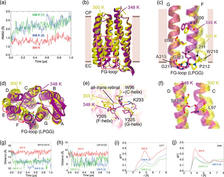
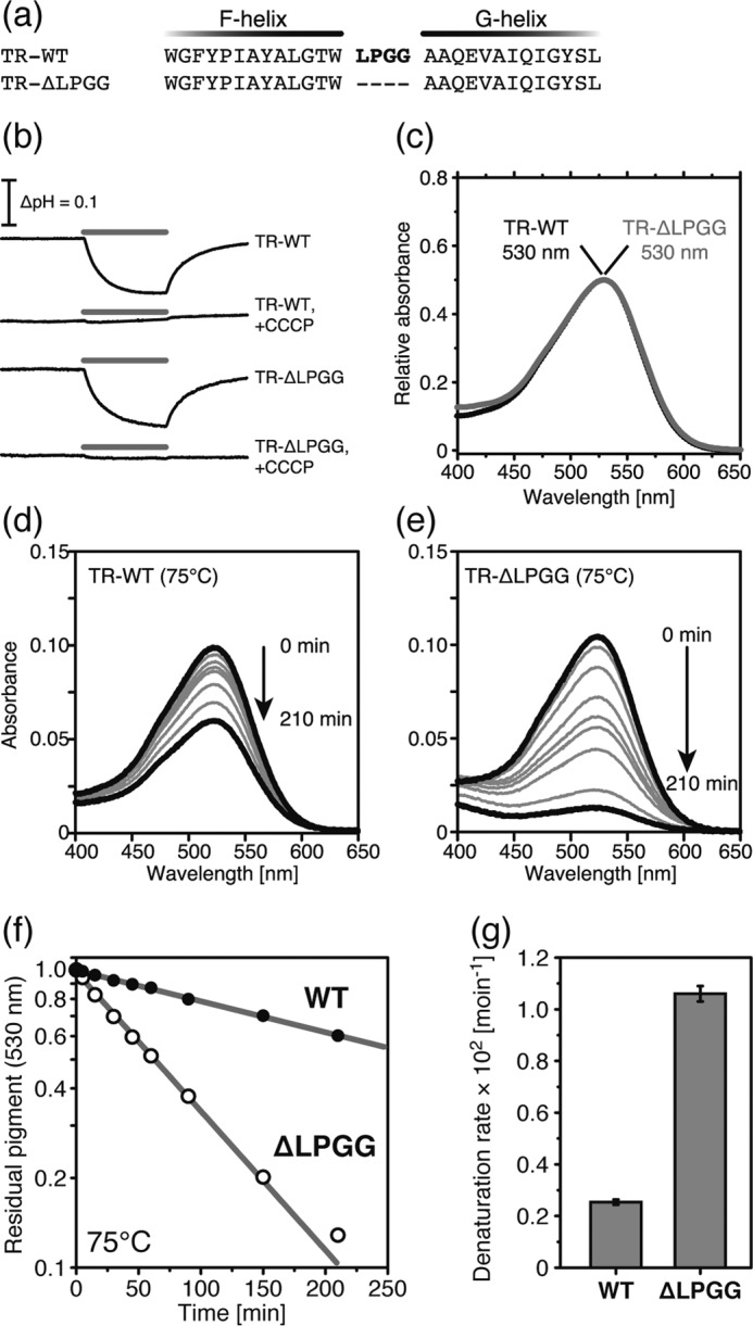
Thermophilic rhodopsin (TR) is a photoreceptor protein with an extremely high thermal stability and the first characterized light-driven electrogenic proton pump derived from the extreme thermophile Thermus thermophilus JL-18. In this study, we confirmed its high thermal stability compared with other microbial rhodopsins and also report the potential availability of TR for optogenetics as a light-induced neural silencer. The x-ray crystal structure of TR revealed that its overall structure is quite similar to that of xanthorhodopsin, including the presence of a putative binding site for a carotenoid antenna; but several distinct structural characteristics of TR, including a decreased surface charge and a larger number of hydrophobic residues and aromatic-aromatic interactions, were also clarified. Based on the crystal structure, the structural changes of TR upon thermal stimulation were investigated by molecular dynamics simulations. The simulations revealed the presence of a thermally induced structural substate in which an increase of hydrophobic interactions in the extracellular domain, the movement of extracellular domains, the formation of a hydrogen bond, and the tilting of transmembrane helices were observed. From the computational and mutational analysis, we propose that an extracellular LPGG motif between helices F and G plays an important role in the thermal stability, acting as a "thermal sensor." These findings will be valuable for understanding retinal proteins with regard to high protein stability and high optogenetic performance.

Keywords: MD simulation; X-ray crystallography; membrane protein; optogenetics; photoreceptor; proton pump; thermal stability.


Figures

FIGURE 1.
FIGURE 1.
Thermal stability and optogenetic availability of TR. a, phylogenetic tree of microbial proton-pumping rhodopsins of known structure. PR, ESR, and ARII stand for proteorhodopsin, Exiguobacterium sibiricum rhodopsin, and Acetabularia rhodopsin II, respectively. b, denaturation kinetics of TR, XR, BR, and AR3 at 75 °C. The broken lines represent fitting curves of a single exponential function. c, denaturation rates at 75 °C estimated from the kinetics shown in b (n = 3). d, expression of TR::GFP and AR3::GFP in transgenic C. elegans by the pan-neuronal promoter. e, paralyzing activity of TR and AR3. Upper panel, elongation of the body length of C. elegans worms expressing TR::GFP in body wall muscle cells (n = 7). Ten pulses of green light (0.8 milliwatt/mm2, 550 nm) of 1.0 s in duration with 1.0-s interstimulus intervals were applied as indicated by the gray bars. Lower panel, elongation of the body length of worms expressing either TR::GFP (n = 4) or AR3::GFP (n = 4) in neurons with 10 green pulses (4.4 milliwatts/mm2, 550 nm) of 1.0 s with 1.0-s interstimulus intervals. f, the average time for the half of the body length change in worms expressing TR and AR3 calculated from the data shown in e, lower panel.
FIGURE 2.
FIGURE 2.
Amino acid sequence alignments of microbial proton-pumping rhodopsins generated by MUSCLE (multiple sequence comparison by log-expectation). Identical residues among these molecules are shown in gray boxes and by asterisks. The transmembrane helices (A-to-G helices), are shown in boxes. The helices belonging to AR3 are not shown because its crystal structure is lacking. The hydrophobic residues of TR and XR are shown in bold blue letters. TR and XR have non-identical aromatic residues, shown in bold red letters, and involve the aromatic-aromatic interactions. The LPGG sequence in the FG loop is shown in a red box. The conserved residue Gly152, which is assumed to contribute to secondary chromophore binding in TR, is highlighted in bold green letters. The residual numbers of TR are shown at the top of the sequence.
FIGURE 3.
FIGURE 3.
Structural details of TR. a, overall structure of TR shown in ribbon representation. Transmembrane α-helices A–G are shown in pink, interhelical loops in the cytoplasmic (CP) and extracellular (EC) sides of the membrane are shown in gray, and β-strands in the BC loop are shown in blue. All-trans-retinal connected to Lys233 via a protonated Schiff base linkage is shown as a yellow stick. b, superimposed structures of TR (pink) and XR (18) (white; PDB ID: 3DDL) and TR and BR (40) (white; PDB ID: 1C3W). r.m.s.d. values were calculated using carbonyl carbon. Magnified views of the retinal chromophore are shown below. c, expected carotenoid-binding cavity in TR. The carotenoid SX in the XR structure (18) is superimposed on the E- and F-helices in the TR structure. The magnified view shows the geometry between SX in XR and the all-trans-retinal in TR. The amino acid sequence alignments of the E- and F-helices are shown at the bottom of the panel. The residues involved in carotenoid binding in XR are conserved in the TR sequence (bold red letters). The most important Gly residue is conserved in TR as Gly152. d, putative proton transport pathway in TR. The retinal and residues involved in proton transport are shown in stick representation.
FIGURE 4.
FIGURE 4.
Structural comparison of TR and XR. a, superimposed structures of TR (pink) and XR (blue) in which the identical and non-identical residues in their amino acid sequence alignments (also shown in Fig. 2) are shown in white and color, respectively. The retinal is shown as a yellow stick. b, table showing the difference in the number of residues and intramolecular interactions of TR and XR. Polar residues: Asn, Gln, Ser, Thr, Asp, Glu, His, Lys, and Arg. Hydrophobic residues: Ala, Phe, Ile, Leu, Met, Val, Trp, Pro, and Tyr. Aromatic residues: Phe, Tyr, and Trp. c and d, aromatic-aromatic interactions of TR and XR. Side chains of the identical and non-identical aromatic residues forming aromatic-aromatic interactions are shown in white and colored spherical representations, respectively. e and f, surface potential distributions of TR and XR. The electrostatic potential surfaces are mapped at contouring levels from −10 kT/e (blue) to 10 kT/e (red).
FIGURE 5.
FIGURE 5.
Temperature-induced conformational changes of TR observed in MD simulations. a, r.m.s.d. values of Cα atoms of TR in MD simulations with respect to the x-ray crystallographic structure at 300 K (solid red line) and at 348 K (dotted green line and dashed blue line). b, overall structures of TR simulated at 300 K (yellow) and 348 K (purple). The upper and lower boxed areas indicate regions where a hydrogen bond between the C- and D-helices is newly formed at 348 K and the extracellular region of the F- and G-helices, respectively. c, extracellular regions of the F- and G-helices simulated at 300 and 348 K. View is from the back side of the protein depicted in a. d, view of the extracellular side of the protein depicted in a. e, the region near the retinal chromophore simulated at 300 and 348 K. f, the region around Leu97 in the C-helix simulated at 300 and 348 K. A hydrogen bond between Leu97 and Ser129 is newly formed at 348 K. g, time evolution of distance between Cα atoms of Trp210 and Ala215, which are located at both ends of the FG loop. h, time evolution of the distance between the Cα atoms of Leu97 and Ser. i, RDF of water molecules from the Cγ atom of Leu211. j, RDF of water molecules from the Cη2 atom of Trp96.
FIGURE 6.
FIGURE 6.
Comparison of TR-WT and TR-ΔLPGG. a, partial amino acid sequence from F- to G-helices. Bold red letters represent the LPGG sequence (Leu211-Pro212-Gly213-Gly21 4) in the extracellular loop between the F- and G-helices. b, light-induced pH changes of TR-WT and TR-ΔLPGG in E. coli suspension in the presence of 100 mm NaCl. The pH was decreased during the 520 ± 10-nm light irradiation for 3 min, shown by the gray bars, in the absence of CCCP. In the presence of 10 μm CCCP, the pH change disappeared. c, visible absorption spectra of TR-WT (black) and TR-ΔLPGG (gray). The absorption maxima were 530 nm. d and e, time-dependent thermal denaturation of TR-WT (d) and TR-ΔLPGG (e) at 75 °C. f, the denaturation kinetics of TR-WT (black) and TR-ΔLPGG (white) at 75 °C. The data were analyzed by a single exponential decay function shown by the gray lines. g, the denaturation rate constants are shown as a bar graph.

References

    1. Ernst O. P., Lodowski D. T., Elstner M., Hegemann P., Brown L. S., and Kandori H. (2014) Microbial and animal rhodopsins: structures, functions, and molecular mechanisms. Chem. Rev. 114, 126–163 - PMC - PubMed
    1. Grote M., Engelhard M., and Hegemann P. (2014) Of ion pumps, sensors and channels: perspectives on microbial rhodopsins between science and history. Biochim. Biophys. Acta 1837, 533–545 - PubMed
    1. Inoue K., Tsukamoto T., and Sudo Y. (2014) Molecular and evolutionary aspects of microbial sensory rhodopsins. Biochim. Biophys. Acta 1837, 562–577 - PubMed
    1. Martinez A., Bradley A. S., Waldbauer J. R., Summons R. E., and DeLong E. F. (2007) Proteorhodopsin photosystem gene expression enables photophosphorylation in a heterologous host. Proc. Natl. Acad. Sci. U.S.A. 104, 5590–5595 - PMC - PubMed
    1. Yoshizawa S., Kawanabe A., Ito H., Kandori H., and Kogure K. (2012) Diversity and functional analysis of proteorhodopsin in marine Flavobacteria. Environ. Microbiol. 14, 1240–1248 - PubMed

Publication types

MeSH terms

LinkOut - more resources