. 2016 Feb 19;351(6275):867-71.
doi: 10.1126/science.aad8282. Epub 2016 Jan 14.

Structures of a CRISPR-Cas9 R-loop complex primed for DNA cleavage

Affiliations

Structures of a CRISPR-Cas9 R-loop complex primed for DNA cleavage

Fuguo Jiang et al. Science. .

Abstract

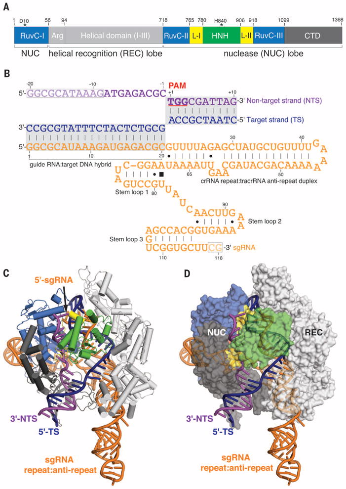
Bacterial adaptive immunity and genome engineering involving the CRISPR (clustered regularly interspaced short palindromic repeats)-associated (Cas) protein Cas9 begin with RNA-guided DNA unwinding to form an RNA-DNA hybrid and a displaced DNA strand inside the protein. The role of this R-loop structure in positioning each DNA strand for cleavage by the two Cas9 nuclease domains is unknown. We determine molecular structures of the catalytically active Streptococcus pyogenes Cas9 R-loop that show the displaced DNA strand located near the RuvC nuclease domain active site. These protein-DNA interactions, in turn, position the HNH nuclease domain adjacent to the target DNA strand cleavage site in a conformation essential for concerted DNA cutting. Cas9 bends the DNA helix by 30°, providing the structural distortion needed for R-loop formation.


Figures

Fig. 1
Fig. 1. Crystal structure of Cas9-sgRNA-dsDNA ternary complex
(A) Domain organization of S. pyogenes Cas9. (B) Schematic diagram of the sgRNA–target DNA complex.The target DNA strand and displaced nontarget strand are colored dark blue and purple, respectively. The PAM sequence is underlined, and the sgRNA sequence is highlighted in orange. Dashes and dots represent canonical and noncanonical Watson-Crick base pairs, respectively. Filled square denotes the base stacking interaction. Dashed boxes outline the portions of sgRNA and DNA that are not visible in the electron density map. (C and D) Ribbon (C) and surface (D) representations of the Cas9-sgRNA-dsDNA structure, color-coded as defined in (A) and (B).
Fig. 2
Fig. 2. Protein contacts that open and kink the nontarget DNA strand
(A) Structural comparison of sgRNA-DNA complexes in existing DNA-bound Cas9 structures (with sgRNA scaffold omitted for clarity). The ordered nontarget DNA strand in the dsDNA-bound structure is shown with a simulated-annealing omit Fo-Fc electron density map contoured at 1.5 σ. The sgRNA-DNA complexes in the ssDNA-bound (PDB ID 4OO8) and PAM-containing partial duplex-bound (PDB ID 4UN3) structures are colored in cyan and beige, respectively. (B) Schematic showing key Cas9-dsDNA interactions. For clarity, only the unwound nontarget strand and the seed RNA–target DNA heteroduplex are shown. Residues specifically interacting with the PAM motif are highlighted in red. Hydrogen bonds or electrostatic interactions are indicated by dashed lines; hydrophobic contacts and van der Waals interactions are shown as solid lines. (C) Position of nontarget strand in RuvC active site. The red sphere depicts the scissile phosphate. Notably, the RuvC domain in the dsDNA-bound structure (marine) superimposes well with that in the Mn2+-bound apo-Cas9 form (gray), with two Mn2+ ions fit snugly in the active center.
Fig. 3
Fig. 3. dsDNA unwinding induces drastic conformational changes in Cas9’s HNH domain and the linker regions connecting it to RuvC domain
(A) Comparison of HNH nuclease domain with that observed in a PAM-containing partial duplex-bound structure. The complexes are aligned through the less-flexible RuvC, helical domain 1, and PAM-interacting C-terminal domain (CTD) and presented in the same direction, but side by side. There is a ~180° rotation (marked by an arrow) of the L1-HNH-L2 region in the dsDNA-bound structure (right) along an axis perpendicular to the central channel relative to that of the partial duplex-bound state (left). (B) Close-up view of the HNH active site and L2 linker. The aromatic ring of Phe916 intercalated between positions −3 and −4 is shown as a stick. (C) Close-up view of the L1 linker.
Fig. 4
Fig. 4. Architecture of the Cas9-R loop complex
(A and C) Cryo-EM reconstructions of sgRNA-bound Cas9 (A) and Cas9-sgRNA bound to a 40-bp target dsDNA (C) at 4.5- and 6.0-Å resolution (using the 0.143 gold standard Fourier Shell Correlation criterion), respectively. Subunits are segmented and colored as indicated. (B) Close-up view of the sgRNA (orange) from our sgRNA-bound structure showing that the 5′ end of the guide (nucleotides 1 to 10, red) undergoes a dramatic kink and is buried within a pocket created by the HNH (light green) and RuvC (light blue) nuclease domains of Cas9. The 5′ end of the guide is reorganized upon base pairing with the target strand in the DNA-bound crystal structure presented in Fig. 1 (blue). (D) R-loop density composed of sgRNA (dark gray) and target DNA (dark blue) and Cas9 (transparent surface), with the HNH domain removed for clarity. (E and F) R-loop density composed of sgRNA (orange) and target DNA (dark blue) with the pseudo-atomic model reveals local bending of the dsDNA by ~30°, creating an angle of 150° between ends (arrow) (E) and large distortions as compared to a linear helical duplex extending from the PAM-proximal end (F).

Comment in

References

    1. Wiedenheft B, Sternberg SH, Doudna JA. Nature. 2012;482:331–338. - PubMed
    1. Sorek R, Lawrence CM, Wiedenheft B. Annu Rev Biochem. 2013;82:237–266. - PubMed
    1. Barrangou R, Marraffini LA. Mol Cell. 2014;54:234–244. - PMC - PubMed
    1. Marraffini LA. Nature. 2015;526:55–61. - PubMed
    1. van der Oost J, Westra ER, Jackson RN, Wiedenheft B. Nat Rev Microbiol. 2014;12:479–492. - PMC - PubMed

Publication types

Associated data