Review
. 2018 Mar 30;293(13):4616-4627.
doi: 10.1074/jbc.TM117.000180. Epub 2018 Feb 6.

Bacterial copper storage proteins

Affiliations
Review

Bacterial copper storage proteins

Christopher Dennison et al. J Biol Chem. .

Abstract

Copper is essential for most organisms as a cofactor for key enzymes involved in fundamental processes such as respiration and photosynthesis. However, copper also has toxic effects in cells, which is why eukaryotes and prokaryotes have evolved mechanisms for safe copper handling. A new family of bacterial proteins uses a Cys-rich four-helix bundle to safely store large quantities of Cu(I). The work leading to the discovery of these proteins, their properties and physiological functions, and how their presence potentially impacts the current views of bacterial copper handling and use are discussed in this review.

Keywords: bacterial copper homeostasis; copper; copper storage; copper transport; metal homeostasis; metalloprotein; methane oxidation; methanotrophs; structural biology.


Conflict of interest statement

The authors declare that they have no conflicts of interest with the contents of this article

Figures

Figure 1.
Figure 1.
Structures of Cu(I)–Mbns and Mbn-mediated copper uptake. A and B, the crystal structures of the Cu(I)–Mbns from M. trichosporium OB3b (A, PDB file 2XJH) (54) and Methylocystis hirsuta CSC1 (B, PDB file 2YGI) (52). Below the structures are the sequences of the leader (black and underlined) and core peptides that make up MbnA. Core peptides are modified to give the Mbn, and the M. hirsuta CSC1 Cu(I)–Mbn structure is of a form with the three C-terminal residues cleaved (amino acids are numbered according to the sequence of the core peptides). The Cu(I) ions are shown as orange spheres ligated by the sulfur atoms (S1 and S2) from thioamide/enethiol groups, and two oxazolone (oxa) ring nitrogens in M. trichosporium OB3b Cu(I)–Mbn, with the N-terminal coordinating heterocycle being a pyrazinediol (pyraA) in M. hirsuta CSC1 Cu(I)–Mbn. Other differences include a sulfate-modified Thr side chain in M. hirsuta CSC1 Cu(I)–Mbn and the overall hairpin-like structure of this Cu(I)–Mbn compared with the more compact M. trichosporium OB3b Cu(I)–Mbn. Also shown are copper uptake by (lines) and relative sMMO activity of (bars) M. trichosporium OB3b (C) and M. hirsuta CSC1 (D) cells after the addition of M. trichosporium OB3b Cu(I)–Mbn (open gray triangles and gray bars) and M. hirsuta CSC1 Cu(I)–Mbn (open cyan circles and cyan bars) to sMMO-active cells. In both cases, copper uptake and switchover from sMMO to pMMO is faster with the native Cu(I)–Mbn (52).
Figure 2.
Figure 2.
Discovery and characterization of Csp1 in M. trichosporium OB3b. A, copper content of anion-exchange soluble extracts of M. trichosporium OB3b purified on a gel-filtration column and SDS-PAGE analysis of the fractions eluted between 8 and 15 ml (35). The intensity profile of the band indicated with an arrow matches that of the main copper peak. B, sequence of this 146 amino acid residue protein (MtCsp1) has a predicted Tat signal peptide (bold) and 13 Cys residues (highlighted yellow) largely present in CXXXC and CXXC motifs (underlined). C, tetrameric arrangement in the crystal structure (PDB file 5FJD) (35) of the overexpressed predicted mature form of MtCsp1 (Gly-1 to Ala-122) with the pink (top left) four-helix bundle monomer (helices numbered), shown in detail (D), highlighting the Cys residues that all point into the core, and residues around the mouth of the bundle, in stick representation. E, structure (PDB file 5FJE) of Cu(I)-MtCsp1 (35) with the metal ions represented as gray spheres and numbered. F, crystal structure (PDB file 1RJU) of a truncated form of S. cerevisiae MT binding eight Cu(I) ions via 10 Cys residues (39) is shown for comparison. G, sequence alignment of the three Csp homologues present in M. trichosporium OB3b created in T-coffee (65) using the predicted mature forms of MtCsp1 and MtCsp2.
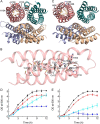
Figure 3.
Figure 3.
Structural and functional studies of Csp3s. A, crystal structure of the apo-MtCsp3 (PDB file 5ARM) tetramer (36). The side chains of the 18 Cys residues pointing into the core and three His residues at the mouth are shown as sticks in the pink (top left) four-helix bundle monomer. The additional small N-terminal α-helix (αN) is labeled in two monomers. B, crystal structure (PDB file 5ARN (36)) of Cu(I)-MtCsp3 (αN omitted) with the metal ions as gray spheres and numbered. C, crystal structure of the apo-BsCsp3 (PDB file 5FIG) tetramer of four-helix bundles (36) using the same representation as in A, with 19 Cys residues pointing into the core of the pink (top left) four-helix bundle monomer. D, growth (37 °C) of ΔcopA E. coli in the absence of (black circles) and plus 1.0 mm (blue circles) Cu(NO3)2 is compared with ΔcopA cells overexpressing BsCsp3 in the absence of (red triangles) and plus 1.0 mm (cyan triangles) Cu(NO3)2 (36). E, growth (37 °C) of WT E. coli in the absence of (black circles) and plus 3.4 mm (blue circles) Cu(NO3)2 is compared with WT cells overexpressing BsCsp3 in the absence of (red triangles) and plus 3.4 mm (cyan triangles) Cu(NO3)2. Overexpressed BsCsp3 can protect ΔcopA (D) and WT (E) E. coli from copper toxicity.
Figure 4.
Figure 4.
Bioinformatics of Csp1s, Csp3s, and the Mbn operon in methanotrophs. The 89 methanotroph genomes currently available in the NCBI database were interrogated with pBLASTp using MtCsp1 and MtCsp3 as search queries. A, Venn diagram of the distribution of Csp1, Csp3, and the Mbn operon (identified by the presence of homologues of M. trichosporium OB3b MbnA, MbnB, and MbnC) in methanotroph genomes. Alignments of 26 and 36 sequences (see Figs. S1 and S2) were used to produce WebLogos (73) for Csp1s (B) and Csp3s (C), respectively. In these, the overall height of the stack at a particular position represents the degree of conservation, whereas the height of the symbol for an amino acid residue (green for polar, purple for neutral, blue for basic, red for acidic, and black for hydrophobic) within the stack signifies relative frequency. Widths are unscaled so less frequently occupied positions in the alignment (see Figs. S1 and S2) are not represented by narrower stacks (composition adjustment was left to the default value for typical amino acid usage). Signal peptides were identified using SignalP (74) and TatP (62), and this region is labeled in B. D, schematic of the model methanotroph M. trichosporium OB3b showing two possible arrangements of the intracytoplasmic membranes that house pMMO and the location and potential roles of MtCsp1, MtCsp2, and MtCsp3. The established cytosolic copper-sensing (CueR) and copper-efflux (CopA) systems, and the known locations and interactions of Mbn (MbnT is a Ton-B-dependent transporter (56) and MbnE is suggested to bind Mbn in the periplasm (58)) are also included. A much more detailed model of copper homeostasis and copper-regulated switchover in a methanotroph can be found in Ref. . This includes the Csps, but it does not discuss their importance.

References

    1. Dennison C. (2005) Investigating the structure and function of cupredoxins. Coord. Chem. Rev. 249, 3025–3054 10.1016/j.ccr.2005.04.021 - DOI
    1. Ridge P. G., Zhang Y., and Gladyshev V. N. (2008) Comparative genomic analyses of copper transporters and cuproproteomes reveal evolutionary dynamics of copper utilization and its link to oxygen. PLoS ONE 3, e1378 10.1371/journal.pone.0001378 - DOI - PMC - PubMed
    1. Turski M. L., and Thiele D. J. (2009) New roles for copper metabolism in cell proliferation, signalling and disease. J. Biol. Chem. 284, 717–721 10.1074/jbc.R800055200 - DOI - PMC - PubMed
    1. Kim B. E., Nevitt T., and Thiele D. J. (2008) Mechanisms for copper acquisition, distribution and regulation. Nat. Chem. Biol. 4, 176–185 10.1038/nchembio.72 - DOI - PubMed
    1. Festa R. A., and Thiele D. J. (2011) Copper: an essential metal in biology. Curr. Biol. 21, R877–R883 10.1016/j.cub.2011.09.040 - DOI - PMC - PubMed

Publication types

MeSH terms

LinkOut - more resources