. 2017 Mar 10;292(10):4022-4033.
doi: 10.1074/jbc.M116.764340. Epub 2017 Jan 18.

Crystallographic Snapshots of Class A β-Lactamase Catalysis Reveal Structural Changes That Facilitate β-Lactam Hydrolysis

Affiliations

Crystallographic Snapshots of Class A β-Lactamase Catalysis Reveal Structural Changes That Facilitate β-Lactam Hydrolysis

Xuehua Pan et al. J Biol Chem. .

Abstract

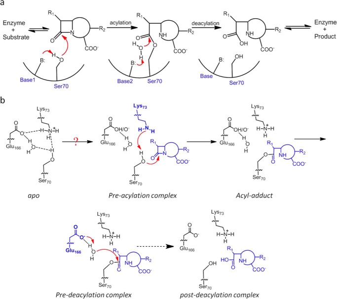
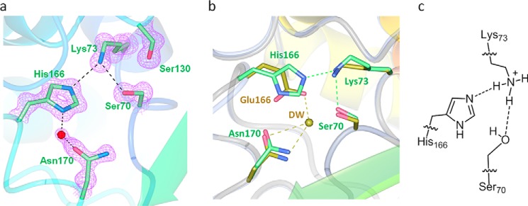
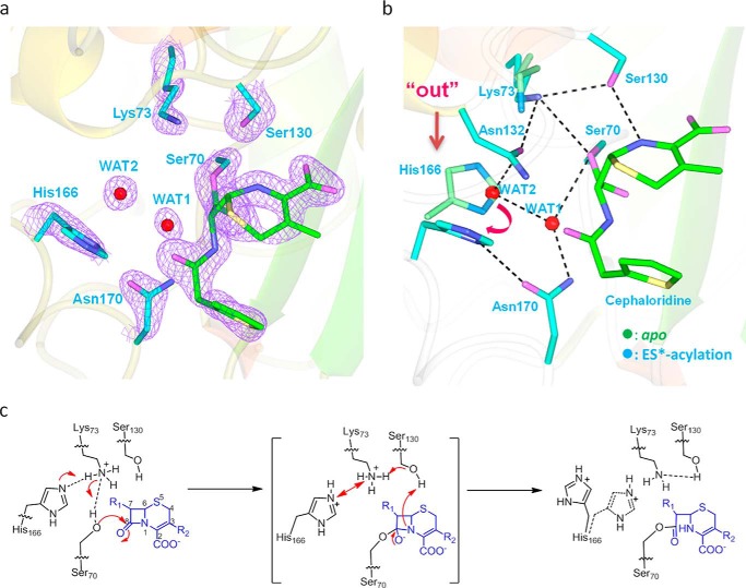
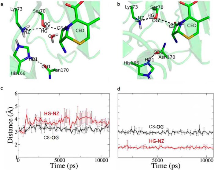
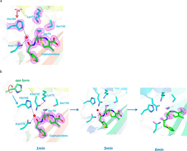
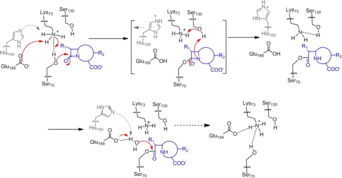
β-Lactamases confer resistance to β-lactam-based antibiotics. There is great interest in understanding their mechanisms to enable the development of β-lactamase-specific inhibitors. The mechanism of class A β-lactamases has been studied extensively, revealing Lys-73 and Glu-166 as general bases that assist the catalytic residue Ser-70. However, the specific roles of these two residues within the catalytic cycle remain not fully understood. To help resolve this, we first identified an E166H mutant that is functional but is kinetically slow. We then carried out time-resolved crystallographic study of a full cycle of the catalytic reaction. We obtained structures that represent apo, ES*-acylation, and ES*-deacylation states and analyzed the conformational changes of His-166. The "in" conformation in the apo structure allows His-166 to form a hydrogen bond with Lys-73. The unexpected "flipped-out" conformation of His-166 in the ES*-acylation structure was further examined by molecular dynamics simulations, which suggested deprotonated Lys-73 serving as the general base for acylation. The "revert-in" conformation in the ES*-deacylation structure aligns His-166 toward the water molecule that hydrolyzes the acyl adduct. Finally, when the acyl adduct is fully hydrolyzed, His-166 rotates back to the "in" conformation of the apo-state, restoring the Lys-73/His-166 interaction. Using His-166 as surrogate, our study identifies distinct conformational changes within the active site during catalysis. We suggest that the native Glu-166 executes similar changes in a less constricted way. Taken together, this structural series improves our understanding of β-lactam hydrolysis in this important class of enzymes.

Keywords: X-ray crystallography; antibiotic resistance; enzyme catalysis; enzyme kinetics; structure-function.


Conflict of interest statement

The authors declare that they have no conflicts of interest with the contents of this article

Figures

FIGURE 1.
FIGURE 1.
Reaction scheme of class A β-lactamase. a, reaction scheme for hydrolysis of β-lactam antibiotics in the active site of class A β-lactamases. B represents the general base. Base1 and Base2 represent different residues for the acylation and deacylation step respectively. b, concerted base model for the acylation step of the catalytic cycle with substrate-induced proton transfer from Lys-73 to Glu-166 so that Lys-73 serves as the general base. The deacylation step employs Glu-166 as the general base. The dashed arrow represents intermediate steps between the pre- and post-deacylation complex.
FIGURE 2.
FIGURE 2.
E166H shows slow deacylation rate and has a distinct pH-dependent profile. a, time-dependent deacylation for E166H-cephaloridine acyl adduct. b, pH dependence of kcat/Km for nitrocefin hydrolysis by E166H mutant β-lactamase.
FIGURE 3.
FIGURE 3.
Active site in the apo structure of E166H reveals a stable Lys-73–His-166 H-bond. a, H-bond interactions involving Lys-73 and His-166 in the active site. FoFc omit map (purple) is drawn in mesh format and contoured at 2.0 σ. The dashed lines indicate hydrogen bonds. b, superposition of E166H active site with that of the wild-type. c, inferred protonation states for Lys-73 and His-166 to sustain the Lys-73–His-166 H-bond.
FIGURE 4.
FIGURE 4.
ES*-acylation structure shows His-166 adopting flipped-out conformation and confirms Lys-73 as general base for acylation. a, active site in the ES*-acylation structure. FoFc omit map (purple) is drawn in mesh format and contoured at 2.0 σ. The dashed lines indicate hydrogen bonds. b, superposition of ES*-acylation (cyan) and apo structures (green). Dashed lines mark the H-bonds. The flipped-out conformation of E166H is marked with an arrow. The tilting of Lys-73 is marked by Lys-73–Ser-130 H-bond. c, scheme of the proposed proton transfer from Lys-73 to His-166 during the acylation step and the resulting “flipped-out” conformation of His-166.
FIGURE 5.
FIGURE 5.
MD simulations to compare two models of the enzyme-substrate complex. Two models of the enzyme-substrate complex were constructed, one with His-166 adopting the “in” conformation as seen in the apo structure and the other with His-166 adopting the “flipped-out” conformation of the ES*-acylation structure. a and c, time course of the distance of Ser-70HG–Lys-73NZ (red) and Ser-70OG-C3 (black) with His-166 adopting “in” conformation. b and d, time course of the distance of Ser-70HG–Lys-73NZ (red) and Ser-70OG-C3 (black) with His-166 adopting flipped-out conformation.
FIGURE 6.
FIGURE 6.
ES* deacylation structure shows “revert-back” conformation of His-166 to serve as exclusive general base for deacylation. The active site of the ES* deacylation structure with “revert-back” conformation for His-166 at the start of the deacylation reaction (a) and tracked by time-dependent X-ray crystallography over a period of 6 min focusing on the disappearance of the acyl adduct (b). FoFc omit map (purple) is drawn in mesh format and contoured at 2.0 σ. The dashed lines indicate hydrogen bonds.
FIGURE 7.
FIGURE 7.
Reaction scheme for wild-type class A β-lactamase after incorporating our findings regarding conformational changes for E166H. PDB codes reported in this work are 5GHX, 5GHY, and 5GHZ.

References

    1. Drawz S. M., and Bonomo R. A. (2010) Three decades of β-lactamase inhibitors. Clin. Microbiol. Rev. 23, 160–201 - PMC - PubMed
    1. Papp-Wallace K. M., Endimiani A., Taracila M. A., and Bonomo R. A. (2011) Carbapenems: past, present, and future. Antimicrob. Agents Chemother. 55, 4943–4960 - PMC - PubMed
    1. Molton J. S., Tambyah P. A., Ang B. S., Ling M. L., and Fisher D. A. (2013) The global spread of healthcare-associated multidrug-resistant bacteria: a perspective from Asia. Clin. Infect. Dis. 56, 1310–1318 - PubMed
    1. Majiduddin F. K., Materon I. C., and Palzkill T. G. (2002) Molecular analysis of β-lactamase structure and function. Int. J. Med. Microbiol. 292, 127–137 - PubMed
    1. Massova I., and Mobashery S. (1998) Kinship and diversification of bacterial penicillin-binding proteins and β-lactamases. Antimicrob. Agents Chemother. 42, 1–17 - PMC - PubMed

Publication types