. 2016 Nov 11;291(46):24054-24064.
doi: 10.1074/jbc.M116.755801. Epub 2016 Sep 28.

Crystal Structure of the Acid Sphingomyelinase-like Phosphodiesterase SMPDL3B Provides Insights into Determinants of Substrate Specificity

Affiliations

Crystal Structure of the Acid Sphingomyelinase-like Phosphodiesterase SMPDL3B Provides Insights into Determinants of Substrate Specificity

Alexei Gorelik et al. J Biol Chem. .

Abstract

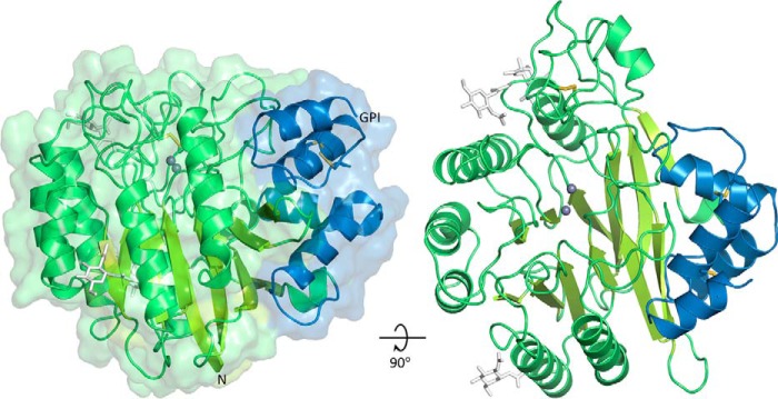
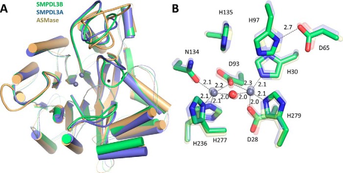
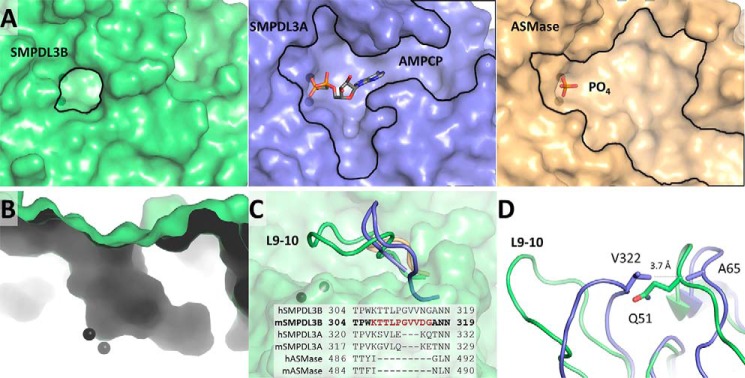
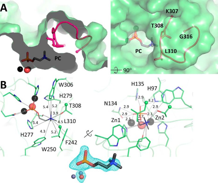
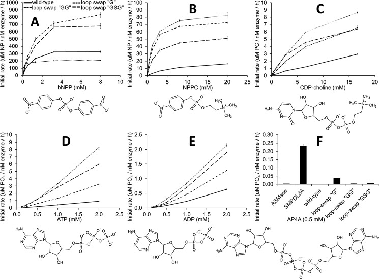
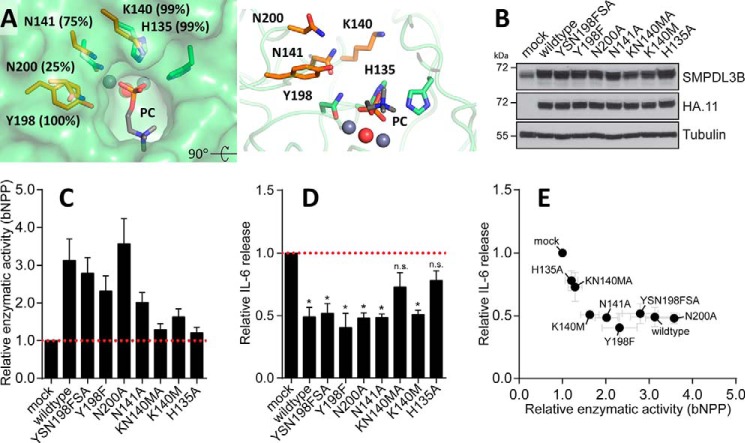
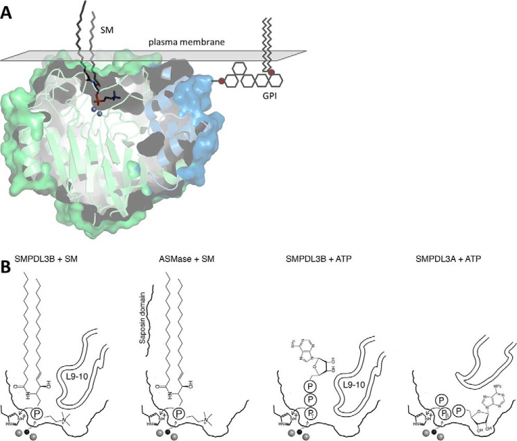
The enzyme acid sphingomyelinase-like phosphodiesterase 3B (SMPDL3B) was shown to act as a negative regulator of innate immune signaling, affecting cellular lipid composition and membrane fluidity. Furthermore, several reports identified this enzyme as an off target of the therapeutic antibody rituximab, with implications in kidney disorders. However, structural information for this protein is lacking. Here we present the high resolution crystal structure of murine SMPDL3B, which reveals a substrate binding site strikingly different from its paralogs. The active site is located in a narrow boot-shaped cavity. We identify a unique loop near the active site that appears to impose size constraints on incoming substrates. A structure in complex with phosphocholine indicates that the protein recognizes this head group via an aromatic box, a typical choline-binding motif. Although a potential substrate for SMPDL3B is sphingomyelin, we identify other possible substrates such as CDP-choline, ATP, and ADP. Functional experiments employing structure-guided mutagenesis in macrophages highlight amino acid residues potentially involved in recognition of endogenous substrates. Our study is an important step toward elucidating the specific function of this poorly characterized enzyme.

Keywords: SMPDL3B; cell surface enzyme; crystal structure; lipid metabolism; sphingomyelinase; substrate specificity.


Figures

FIGURE 1.
FIGURE 1.
Crystal structure of SMPDL3B. The catalytic domain is shown in green, and the C-terminal subdomain is in blue. Zinc ions are represented by gray spheres. N-Linked glycans (white sticks) are only partially displayed for clarity. Disulfide bonds are shown as yellow sticks. The N terminus and the putative C-terminal ω-site of GPI anchor attachment are indicated.
FIGURE 2.
FIGURE 2.
Structural comparison of SMPDL3B, SMPDL3A, and ASMase. A, superimposition of the ASMase family of proteins. SMPDL3B is shown in green, SMPDL3A is in blue (PDB code 5FC6), and ASMase is in yellow (PDB code 5HQN). Loops on the face of the protein containing the active site that diverge between the family members are displayed as thick lines. Loop L9–10 of SMPDL3B (described later) is marked with an asterisk. SMPDL3B shares 28% identity with ASMase (root mean square deviation of 0.851 Å over 284 alpha carbons) and 38% identity with SMPDL3A (root mean square deviation of 0.606 Å over 295 alpha carbons). The saposin domain of ASMase is not shown. B, comparison of the SMPDL3B active site with that of SMPDL3A and ASMase. Active site residues of SMPDL3B are shown as green sticks. The active site of SMPDL3A (transparent blue sticks) and ASMase (transparent yellow sticks) are overlaid. Zinc ions are represented by gray spheres. The bridging water molecule is displayed as a red sphere. Distances are indicated (Å).
FIGURE 3.
FIGURE 3.
Region around active site. A, the pocket at the active site of SMPDL3B (green). Zinc ions are represented by black spheres. The substrate binding site of SMPDL3A in complex with a nucleotide (blue, PDB code 5FC6) and ASMase bound to phosphate (yellow, PDB code 5HQN) are shown for comparison. B, boot-shaped cavity at the active site of SMPDL3B. C, the loop L9–10 of SMPDL3B (green) is compared with the corresponding loops of SMPDL3A (blue) and ASMase (yellow). Sequences of the loops of human and mouse homologs are aligned. D, conformational differences between the L9–10 loop of SMPDL3B and SMPDL3A are highlighted.
FIGURE 4.
FIGURE 4.
Phosphocholine bound in the active site of SMPDL3B. A, PC (sticks) in the active site of SMPDL3B, in proximity to loop L9–10 (pink). B, residues that interact with the choline group of PC are shown as sticks (left). Distances between the choline nitrogen atom and the closest protein non-hydrogen atoms are indicated (Å). Residues that form contacts with the phosphate portion of PC, including zinc ions and water, are shown as sticks (right). Interatomic distances are indicated (Å). The electron density FoFc simulated annealing omit map around PC, and the nucleophilic water molecule is contoured at 3σ.
FIGURE 5.
FIGURE 5.
In vitro enzymatic activity against various substrates. A–F, the activity profiles of purified SMPDL3B are shown for the wild type protein (solid line), as well as for loop swaps of L9–10 by glycine, GG, or GSG (dashed lines). The chemical structures of substrates are depicted. In F, activity of purified murine ASMase (24) and SMPDL3A (22) is also shown. The values are the means and standard deviations of triplicates representative of one of two experiments.
FIGURE 6.
FIGURE 6.
Functional impact of SMPDL3B mutants on LPS-induced IL-6 release in macrophages. A, residues lining the wall of the channel leading to the active site are displayed in orange along with their degree of conservation in vertebrates. Other adjacent amino acids, including the catalytic machinery, are in green. B, Western blot analysis for SMPDL3B, HA, and tubulin in samples from RAW264.7 macrophages stably transduced with an empty vector (mock) or HA-tagged wild type or mutant murine SMPDL3B constructs as indicated. C and D, RAW264.7 macrophages stably transduced with an empty vector (mock) or HA-tagged wild type or mutant murine SMPDL3B constructs were stimulated with 100 ng/ml LPS for 8 h. The cells were probed for enzymatic activity by incubation with bNPP (C), and supernatants were analyzed for IL-6 release by ELISA (D). Experimental data were normalized to mock and represent means ± S.E. from five independent biological replicates. Statistical significance was tested using a one sample t test against a hypothetical mean value of 1. *, p ≤ 0.05; n.s., not significant. E, relative enzymatic activity and relative IL-6 release shown in C and D plotted against each other.
FIGURE 7.
FIGURE 7.
Proposed binding modes of potential substrates. A, SM (dark gray sticks) was manually placed in the substrate binding site using bound PC as a guide. A GPI anchor (stylized gray lines) is attached to the putative C-terminal ω-site. The surface of the plasma membrane is represented by a gray plane. B, comparison of potential substrate positioning in SMPDL3B and ASMase for SM and in SMPDL3B and SMPDL3A for nucleotide triphosphates. Zinc ions (gray spheres) and the nucleophilic water molecule (black sphere) are shown.

References

    1. Slotte J. P. (2013) Biological functions of sphingomyelins. Prog. Lipid Res. 52, 424–437 - PubMed
    1. Castro B. M., Prieto M., and Silva L. C. (2014) Ceramide: a simple sphingolipid with unique biophysical properties. Prog. Lipid Res. 54, 53–67 - PubMed
    1. Wu J., Hansen G. H., Nilsson A., and Duan R. D. (2005) Functional studies of human intestinal alkaline sphingomyelinase by deglycosylation and mutagenesis. Biochem. J. 386, 153–160 - PMC - PubMed
    1. Airola M. V., and Hannun Y. A. (2013) Sphingolipid metabolism and neutral sphingomyelinases. Handb. Exp. Pharmacol. 215, 57–76 - PMC - PubMed
    1. Kornhuber J., Rhein C., Müller C. P., and Mühle C. (2015) Secretory sphingomyelinase in health and disease. Biol. Chem. 396, 707–736 - PubMed

MeSH terms

Substances