Review
. 2020 Jul 17;295(29):9823-9837.
doi: 10.1074/jbc.REV120.010181. Epub 2020 May 14.

How repertoire data are changing antibody science

Affiliations
Review

How repertoire data are changing antibody science

Claire Marks et al. J Biol Chem. .

Abstract

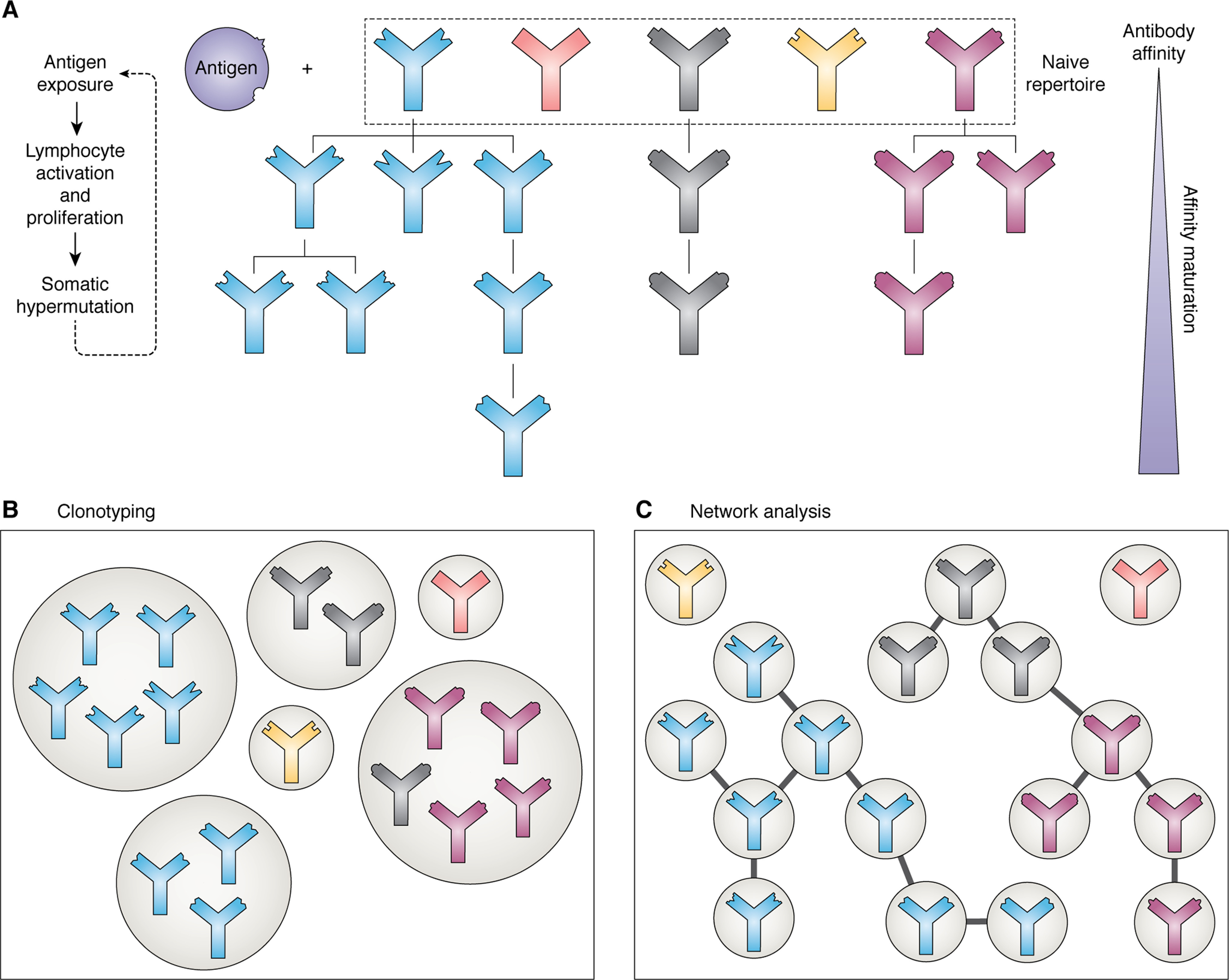
Antibodies are vital proteins of the immune system that recognize potentially harmful molecules and initiate their removal. Mammals can efficiently create vast numbers of antibodies with different sequences capable of binding to any antigen with high affinity and specificity. Because they can be developed to bind to many disease agents, antibodies can be used as therapeutics. In an organism, after antigen exposure, antibodies specific to that antigen are enriched through clonal selection, expansion, and somatic hypermutation. The antibodies present in an organism therefore report on its immune status, describe its innate ability to deal with harmful substances, and reveal how it has previously responded. Next-generation sequencing technologies are being increasingly used to query the antibody, or B-cell receptor (BCR), sequence repertoire, and the amount of BCR data in public repositories is growing. The Observed Antibody Space database, for example, currently contains over a billion sequences from 68 different studies. Repertoires are available that represent both the naive state (i.e. antigen-inexperienced) and that after immunization. This wealth of data has created opportunities to learn more about our immune system. In this review, we discuss the many ways in which BCR repertoire data have been or could be exploited. We highlight its utility for providing insights into how the naive immune repertoire is generated and how it responds to antigens. We also consider how structural information can be used to enhance these data and may lead to more accurate depictions of the sequence space and to applications in the discovery of new therapeutics.

Keywords: B-cell receptor (BCR); adaptive immunity; antibody; bioinformatics; immunology; next-generation sequencing; observed antibody space database; protein sequence; protein structure; structural annotation.


Conflict of interest statement

Conflict of interest—The authors declare that they have no conflicts of interest with the contents of this article.

Figures

Figure 1.
Figure 1.
A, antibody structure. An antibody is made up of four chains: two light (orange) and two heavy (blue). Each chain is made up of a series of domains—the variable domains of the light and heavy chains together are known as the Fv region (shown on the right; PDB entry 12E8). The Fv features six loops known as CDRs (shown in dark blue); these are mainly responsible for antigen binding. B, example sequences for the VH and VL, highlighting the CDR regions and the genetic composition.
Figure 2.
Figure 2.
The cumulative growth of publicly available (redundant) antibody sequences over time (data from the Observed Antibody Space database (28)).
Figure 3.
Figure 3.
The process of affinity maturation and methods of analyzing the resulting antibody repertoires. A, upon exposure to an antigen, those antibodies present in the naive repertoire that are able to bind to it proliferate, undergoing somatic hypermutation to produce variations upon the initial binder. Successive rounds of this process produce antibodies with high affinity. B, clonotyping groups antibodies in the repertoire based on sequence similarity; normally they must originate from the same V and J genes and have an H3 sequence identity of 80–100%. Antibodies of the same clonotype are predicted to bind to the same epitope. C, network analysis of antibody repertoires, where each node is a different sequence and edges are present between them if they meet set sequence similarity criteria. The lineages of different antibodies can be inferred using this method.
Figure 4.
Figure 4.
Sequence is not always a reliable indicator of structural similarity. A, L1 loops of the PDB entries 3PHO (red) and 3QUM (blue). The two loops differ in sequence at every position except one (sequence identity = 10%); however, they have very similar conformations (RMSD = 0.60 Å). B, H3 loops of the PDB entries 5I1G (red) and 5I1C (blue). These loops have very similar sequences (sequence identity = 92%) and therefore may be predicted to have similar structures; however, this is not the case (RMSD = 4.15 Å). RMSDs were calculated across all backbone atoms after superposition of the anchor residues (two residues on each side of the loop, shown in gray).
Figure 5.
Figure 5.
WebLogo representations for the second framework region (residues 39–55 in the IMGT numbering scheme) for known human and mouse antibody sequences. Hydrophobic amino acids are shown in red, hydrophilic in blue, and neutral in gray. Data were extracted from OAS; we only considered repertoires from individuals with no disease and no vaccine recorded. Whereas amino acid usage is the same at many positions along the sequence, it can be seen that there are differences that could potentially be used to measure “humanness” and guide the humanization process. For example, it is rare to observe lysine at position 43 in human antibodies, but this is common in mice. Changing a lysine to an arginine at this position in a potential therapeutic may therefore reduce immunogenicity.

References

    1. Sela-Culang I., Kunik V., and Ofran Y. (2013) The structural basis of antibody-antigen recognition. Front. Immunol. 4, 302 10.3389/fimmu.2013.00302 - DOI - PMC - PubMed
    1. Saper C. B. (2009) A guide to the perplexed on the specificity of antibodies. J. Histochem. Cytochem. 57, 1–5 10.1369/jhc.2008.952770 - DOI - PMC - PubMed
    1. Ecker D. M., Jones S. D., and Levine H. L. (2015) The therapeutic monoclonal antibody market. mAbs 7, 9–14 10.4161/19420862.2015.989042 - DOI - PMC - PubMed
    1. Raybould M. I. J., Marks C., Lewis A. P., Shi J., Bujotzek A., Taddese B., and Deane C. M. (2020) Thera-SAbDab: the therapeutic structural antibody database. Nucleic Acids Res. 48, D383–D388 10.1093/nar/gkz827 - DOI - PMC - PubMed
    1. Kaplon H., and Reichert J. M. (2019) Antibodies to watch in 2019. mAbs 11, 219–238 10.1080/19420862.2018.1556465 - DOI - PMC - PubMed

LinkOut - more resources