. 2019 Nov 15;294(46):17339-17353.
doi: 10.1074/jbc.RA119.009523. Epub 2019 Sep 26.

A carbohydrate-binding family 48 module enables feruloyl esterase action on polymeric arabinoxylan

Affiliations

A carbohydrate-binding family 48 module enables feruloyl esterase action on polymeric arabinoxylan

Jesper Holck et al. J Biol Chem. .

Abstract

Feruloyl esterases (EC 3.1.1.73), belonging to carbohydrate esterase family 1 (CE1), hydrolyze ester bonds between ferulic acid (FA) and arabinose moieties in arabinoxylans. Recently, some CE1 enzymes identified in metagenomics studies have been predicted to contain a family 48 carbohydrate-binding module (CBM48), a CBM family associated with starch binding. Two of these CE1s, wastewater treatment sludge (wts) Fae1A and wtsFae1B isolated from wastewater treatment surplus sludge, have a cognate CBM48 domain and are feruloyl esterases, and wtsFae1A binds arabinoxylan. Here, we show that wtsFae1B also binds to arabinoxylan and that neither binds starch. Surface plasmon resonance analysis revealed that wtsFae1B's Kd for xylohexaose is 14.8 μm and that it does not bind to starch mimics, β-cyclodextrin, or maltohexaose. Interestingly, in the absence of CBM48 domains, the CE1 regions from wtsFae1A and wtsFae1B did not bind arabinoxylan and were also unable to catalyze FA release from arabinoxylan. Pretreatment with a β-d-1,4-xylanase did enable CE1 domain-mediated FA release from arabinoxylan in the absence of CBM48, indicating that CBM48 is essential for the CE1 activity on the polysaccharide. Crystal structures of wtsFae1A (at 1.63 Å resolution) and wtsFae1B (1.98 Å) revealed that both are folded proteins comprising structurally-conserved hydrogen bonds that lock the CBM48 position relative to that of the CE1 domain. wtsFae1A docking indicated that both enzymes accommodate the arabinoxylan backbone in a cleft at the CE1-CBM48 domain interface. Binding at this cleft appears to enable CE1 activities on polymeric arabinoxylan, illustrating an unexpected and crucial role of CBM48 domains for accommodating arabinoxylan.

Keywords: arabinoxylan; carbohydrate esterase family 1; carbohydrate-binding module; crystal structure; enzyme catalysis; enzyme mechanism; ferulic acid esterase; molecular docking; molecular dynamics; structure–function.


Conflict of interest statement

J. B. and K. B. R. M. K. are employees at Novozymes A/S, a company that produces and sells enzymes and microbes for industrial uses

Figures

Figure 1.
Figure 1.
wtsFae1B substrate interactions. A, adsorption analysis of insoluble wheat arabinoxylan binding to wtsFae1B. B, surface plasmon resonance analysis of xylohexaose binding to wtsFae1B.
Figure 2.
Figure 2.
Overall structures of wtsFae1A and wtsFae1B dimers and electron density at the linker regions. A, two-domain structure of wtsFae1A chain A (cyan) and wtsFae1A chain B (green cyan); B, two-domain structure of wtsFae1B chain A (green) and wtsFae1B chain B (lime). The catalytic triads, Ser242–His325–Glu296 for wtsFae1A and Ser272–His325–Asp339 for wtsFae1B, are highlighted in red. Composite omit map is contoured to 1.0σ in blue mesh with a cutoff at 1.6 Å for C, the linker region of wtsFae1A chain A, and D, the linker region of wtsFae1B chain A. The composite omit map was calculated using Phenix with the dataset for PDB codes 6RZO and 6RZN.
Figure 3.
Figure 3.
Hydrogen bonds keeping the CE1 and CBM48 domains in the correct orientation. A, structurally-conserved residues involved in hydrogen bonds forming the rigid wtsFae1A (cyan) and wtsFae1B (green) structures (hydrogen bonds are shown as yellow dashed lines with their length given in Å, and the residues involved are shown as sticks). B, multiple alignment of CE1–CBM48 homologs (see Fig. S6 for complete alignment). The asterisks indicate the residues involved in hydrogen bonds keeping the CE1 and CBM48 domains in the correct relative orientation (wtsFae1B above and wtsFae1A below the multiple alignment). The protein sequences are identified by their GenBankTM accession numbers. The multiple alignment is visualized using ESPript 3.0 (57).
Figure 4.
Figure 4.
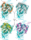
Comparison of wtsFae1A chain A to other CE1 feruloyl esterases. Superimpositions of wtsFae1A chain A (cyan) are shown. A, AmFae1A from A. mucronatus (PDB code 5CXX) (orange); B, BiFae1A from B. intestinalis (PDB code 5VOL) (gray); C, Ets1E from B. proteoclasticus (PDB code 2WTM) (yellow); and D, XynX from H. thermocellum (PDB code 1JJF) (purple). The active site of wtsFae1A is indicated as are the flexible loops and clamps that form a lid on the active-site pocket.
Figure 5.
Figure 5.
Flexibility of Cα in wtsFae1A. Root mean square fluctuation as a function of Cα in wtsFae1A chain A during a 200-ns molecular dynamics simulation is shown.
Figure 6.
Figure 6.
Comparison of the loops forming the lid on the ferulic acid–binding pocket. Multiple alignment shows the loops forming a lid on the ferulic acid–binding pocket. The lid-forming loops/β-clamp are highlighted by green boxes. Asterisk indicates the catalytic residues. The residue numbering refers to wtsFae1A. The sequences were obtained from PDB code 1JJF (H. thermocellum), PDB code 5CXX (A. mucronatus), PDB code 5VOL (B. intestinalis), and PDB code 2WTM (B. proteoclasticus) (see Fig. S2 for complete alignment). The multiple alignment is visualized using ESPript 3.0 (57).
Figure 7.
Figure 7.
wtsFae1A substrate interaction. A, schematic drawing of XA5f2X (xylopyranosyl moieties, black; arabinofuranosyl moiety, green; ferulic acid, purple), and B, wtsFae1A chain A (cyan) docked to XA5f2X (yellow) and ferulic acid from H. thermocellum XynZ (PDB code 1JT2) (white) superimposed. wtsFae1A chain residues interacting directly with XA5f2X and Trp157 are labeled, and hydrogen bonds are shown as dotted lines (yellow), and their length is given in Å.
Figure 8.
Figure 8.
Comparison of wtsFae1A and wtsFae1B substrate-binding cleft and ferulic acid–binding pocket. A, electrostatic plot of wtsFae1A with docked XA5f2X (yellow); B, electrostatic plot of wtsFae1B; C, superimposition of wtsFae1A (cyan) and wtsFae1B (green) surface representations with docked XA5f2X (yellow); D, hydrophobicity plot of wtsFae1A (increase in the red intensity equals increase in hydrophobicity) with docked XA5f2X (yellow); and E, hydrophobicity plot of wtsFae1B (increase in the red intensity equals increase in hydrophobicity).
Figure 9.
Figure 9.
CBM48 structural comparison. A, superimposition of the wtsFae1A (cyan) and wtsFae1B (green) CBM48s and the A. thaliana starch phosphatase Starch Excess4 CBM48 (pink) (PDB code 4PYH); B, superimposition of wtsFae1A (cyan) with docked XA5f2X (yellow) and A. thaliana starch phosphatase Starch Excess4 (pink) in complex with maltohexaose (black); C, as B with the surface of Starch Excess4 presented; D, as B with the surface of wtsFae1A presented.
Figure 10.
Figure 10.
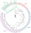
Related starch-binding and carbohydrate-binding domains' relation to CE1-appended CBMs. Phylogenetic tree of the CBM20 (blue), CBM48 (red), CBM69 (purple), and wtsFae1A and wtsFae1B CBM48 homologs (green) is shown. wtsFae1A and wtsFae1B CBM482 are in black. Cladogram highlighting the relative position of protein sequences identified by a reference to GenBankTM or Uniprot accession number and bootstraps values are shown at the nodes. The alignment used to construct the phylogenetic tree is shown in Fig. S8. The phylogenetic tree is visualized using iTOL (58).

References

    1. Naidu D. S., Hlangothi S. P., and John M. J. (2018) Bio-based products from xylan: a review. Carbohydr. Polym. 179, 28–41 10.1016/j.carbpol.2017.09.064 - DOI - PubMed
    1. Garg S. (2016) Xylanase: applications in biofuel production. Curr. Metabolomics 4, 23–37 10.2174/2213235X03666150915211224 - DOI
    1. González-García S., Morales P. C., and Gullón B. (2018) Estimating the environmental impacts of a brewery waste-based biorefinery: bio-ethanol and xylooligosaccharides joint production case study. Ind. Crops Prod. 123, 331–340 10.1016/j.indcrop.2018.07.003 - DOI
    1. Scheller H. V., and Ulvskov P. (2010) Hemicelluloses. Annu. Rev. Plant Biol. 61, 263–289 10.1146/annurev-arplant-042809-112315 - DOI - PubMed
    1. Naran R., Chen G., and Carpita N. C. (2008) Novel rhamnogalacturonan I and arabinoxylan polysaccharides of flax seed mucilage. Plant Physiol. 148, 132–141 10.1104/pp.108.123513 - DOI - PMC - PubMed

Publication types

MeSH terms