. 2019 May 21;116(21):10366-10371.
doi: 10.1073/pnas.1900441116. Epub 2019 May 9.

Effects of α-tubulin acetylation on microtubule structure and stability

Affiliations

Effects of α-tubulin acetylation on microtubule structure and stability

Lisa Eshun-Wilson et al. Proc Natl Acad Sci U S A. .

Abstract

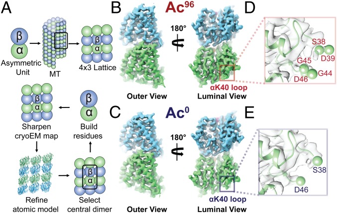
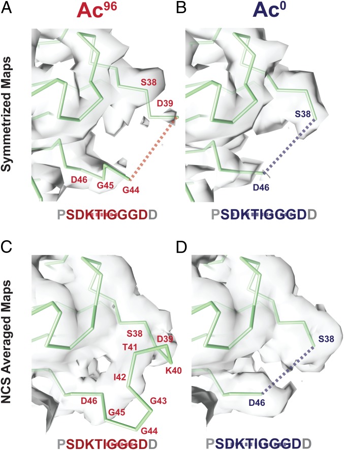
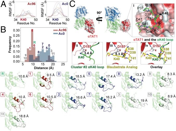
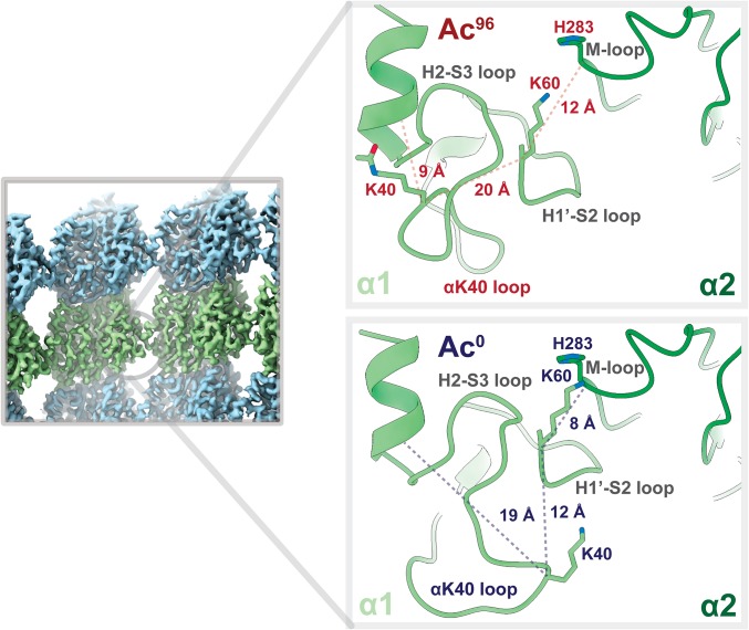
Acetylation of K40 in α-tubulin is the sole posttranslational modification to mark the luminal surface of microtubules. It is still controversial whether its relationship with microtubule stabilization is correlative or causative. We have obtained high-resolution cryo-electron microscopy (cryo-EM) reconstructions of pure samples of αTAT1-acetylated and SIRT2-deacetylated microtubules to visualize the structural consequences of this modification and reveal its potential for influencing the larger assembly properties of microtubules. We modeled the conformational ensembles of the unmodified and acetylated states by using the experimental cryo-EM density as a structural restraint in molecular dynamics simulations. We found that acetylation alters the conformational landscape of the flexible loop that contains αK40. Modification of αK40 reduces the disorder of the loop and restricts the states that it samples. We propose that the change in conformational sampling that we describe, at a location very close to the lateral contacts site, is likely to affect microtubule stability and function.

Keywords: MD; acetylation; cryo-EM; microtubule; tubulin modifications.


Conflict of interest statement

Conflict of interest statement: E.N. and C.J. were coauthors in the 2016 review article “Microtubules: 50 Years on from the discovery of tubulin” [Borisy G, et al. (1)].

Figures

Fig. 1.
Fig. 1.
High-resolution maps of 96% acetylated (Ac96) and <1% acetylated (Ac0) MTs. (A) Schematic of the model-building and refinement process in PHENIX. We sharpened a representative 4 × 3 lattice, refined the corresponding atomic structure (3JAR) into our map, and extracted out the central dimer to build additional residues into the αK40 loop. We performed this process iteratively for both the Ac96 and Ac0. The structure of the Ac96 (B) and Ac0 (C) αβ-tubulin heterodimers, respectively, are shown from the outer and luminal views with close-ups of αK40 loop in each state (D and E) low-pass filtered to 3.7 Å.
Fig. 2.
Fig. 2.
Symmetrized and NCS-averaged C1 maps of Ac96 and Ac0 MTs reveal the αK40 loop is more ordered in the Ac96 state. Close-up views of the αK40 loop (P37–D47) in the (A) Ac96 and (B) Ac0 states in the symmetrized maps low-pass filtered to 4 Å and the (C) Ac96 and (D) Ac0 states in the NCS-averaged C1 maps low-pass filtered to 4 Å. The dotted lines indicate missing residues.
Fig. 3.
Fig. 3.
Acetylation restricts the motion and alters the conformational ensemble of the αK40 loop. (A) Per-residue root-mean-square fluctuation (RMSF) analyses were determined over the course of 12 ns for residues 34–50 the C1 maps using GROMACs in PLUMED and graphed using the MDAnalysis. The different colored lines refer to the eight different replicas. (B) Ensemble modeling of the loop across Ac96 and Ac0 states using density restrained MD. Frames were classified into 1 of 11 clusters by conformation. Clusters either had a greater number of Ac96 frames (red), Ac0 frames (blue), or an equal number of frames from both states (gray). The reference is shown in green. The unique conformations of each of the 12 clusters (0–11) are shown below. (C) αTAT1 cocrystalized with a bisubstrate analog consisting of α-tubulin residues 38–41 (PDB ID code 4PK3) is shown bound to αβ-tubulin sampling the αK40 loop of cluster 2 from the metainference ensemble. αTAT1 is colored by hydrophobicity, where hydrophobic regions are red. (C, i) After backbone alignment of the surrounding residues of the bisubstrate analog, αK40 from cluster 2 fits directly into the catalytic groove, highlighted by the black arrow. (C, iiiv) Similar to the bisubstrate analog (shown in yellow), αK40 from the cluster #2 loop (shown in green) is positioned very close to αTAT1:D157, which stabilizes the enzyme–tubulin interface.
Fig. 4.
Fig. 4.
Acetylation may weaken lateral interactions. Close-up view of the lateral contacts between two α-tubulin monomers at a nonseam location (α1, light green; α2, dark green). K40 in α1 of the Ac0 state is 8 Å closer to the M-loop of α2 and appears to buttress the H1′–S2 loop, providing support for the vital α1K60–α2H283 lateral interaction. By contrast, that support is lost in the Ac96 state because the acetylated K40 now packs much closer to the hydrophobic, inner core.

References

    1. Borisy G, et al. (2016) Microtubules: 50 years on from the discovery of tubulin. Nat Rev Mol Cell Biol 17:322–328. - PMC - PubMed
    1. Nogales E, Whittaker M, Milligan RA, Downing KH (1999) High-resolution model of the microtubule. Cell 96:79–88. - PubMed
    1. Zhang R, Alushin GM, Brown A, Nogales E (2015) Mechanistic origin of microtubule dynamic instability and its modulation by EB proteins. Cell 162:849–859. - PMC - PubMed
    1. Mitchison TJ. (1993) Localization of an exchangeable GTP binding site at the plus end of microtubules. Science 261:1044–1047. - PubMed
    1. Mitchison T, Kirschner M (1984) Dynamic instability of microtubule growth. Nature 312:237–242. - PubMed

Publication types