. 2018 Jan 2;215(1):63-75.
doi: 10.1084/jem.20170869. Epub 2017 Nov 22.

Rare PfCSP C-terminal antibodies induced by live sporozoite vaccination are ineffective against malaria infection

Affiliations

Rare PfCSP C-terminal antibodies induced by live sporozoite vaccination are ineffective against malaria infection

Stephen W Scally et al. J Exp Med. .

Abstract

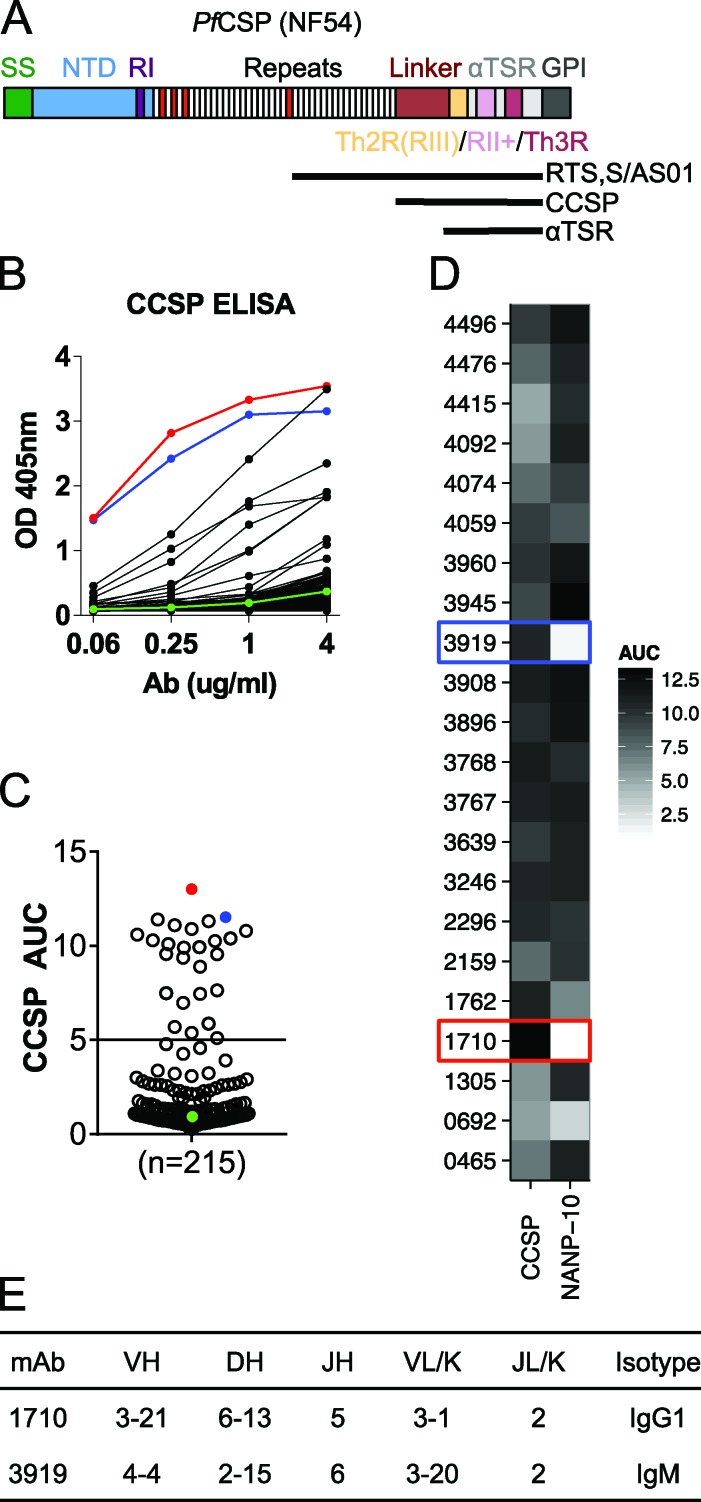
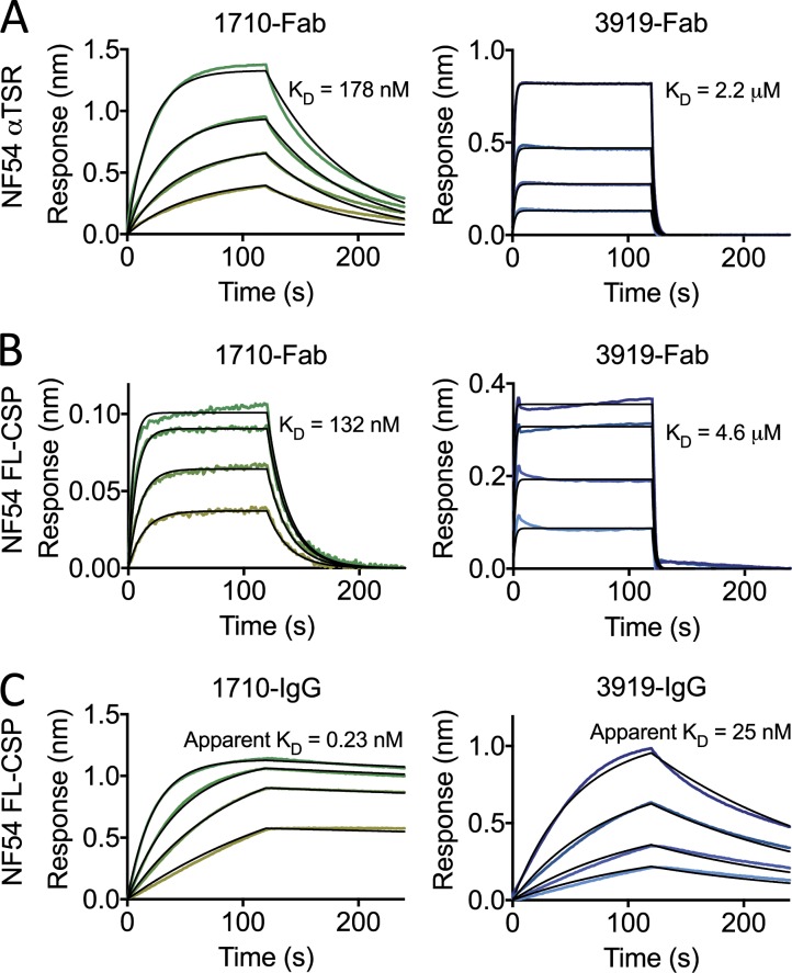
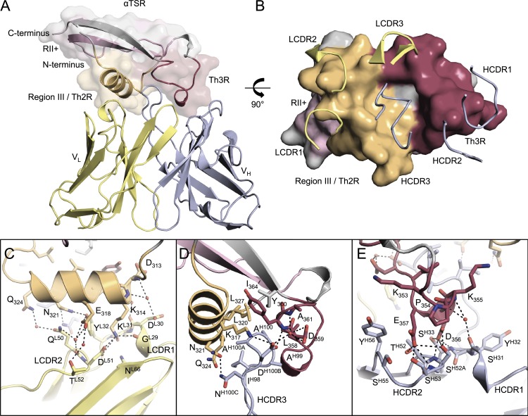
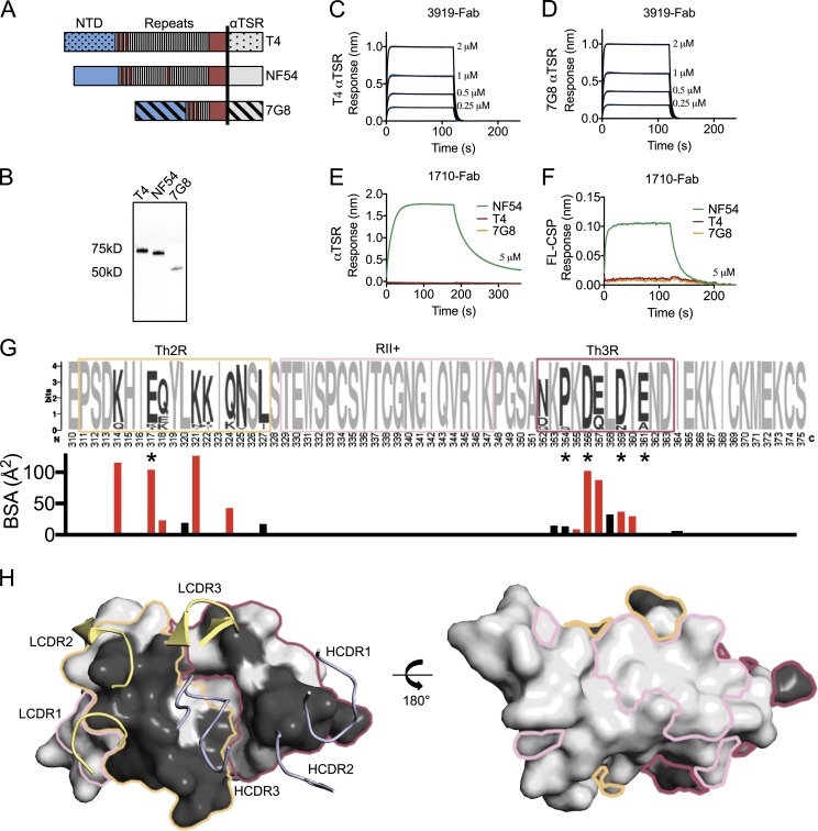
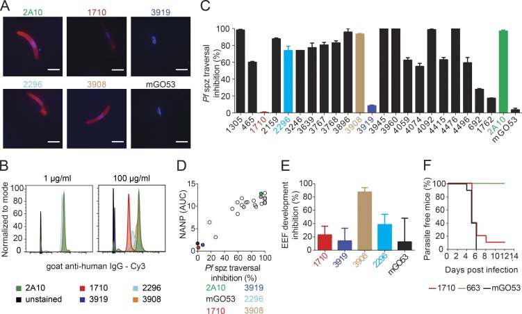
Antibodies against the central repeat of the Plasmodium falciparum (Pf) circumsporozoite protein (CSP) inhibit parasite activity and correlate with protection from malaria. However, the humoral response to the PfCSP C terminus (C-PfCSP) is less well characterized. Here, we describe B cell responses to C-PfCSP from European donors who underwent immunization with live Pf sporozoites (PfSPZ Challenge) under chloroquine prophylaxis (PfSPZ-CVac), and were protected against controlled human malaria infection. Out of 215 PfCSP-reactive monoclonal antibodies, only two unique antibodies were specific for C-PfCSP, highlighting the rare occurrence of C-PfCSP-reactive B cells in PfSPZ-CVac-induced protective immunity. These two antibodies showed poor sporozoite binding and weak inhibition of parasite traversal and development, and did not protect mice from infection with PfCSP transgenic Plasmodium berghei sporozoites. Structural analyses demonstrated that one antibody interacts with a polymorphic region overlapping two T cell epitopes, suggesting that variability in C-PfCSP may benefit parasite escape from humoral and cellular immunity. Our data identify important features underlying C-PfCSP shortcomings as a vaccine target.


Figures

Figure 1.
Figure 1.
Characterization of antibodies elicited by immunization with live sporozoites under chemoprophylaxis (PfSPZ-CVac) against C-PfCSP. (A) Schematic representation of FL PfCSP (NF54). PfCSP contains a signal sequence (SS), N-terminal domain (NTD), region I (RI), a tandem repeat region consisting of NANP (white) or NVDP (red) repeats, a linker region, and an α1-helix thrombospondin type-1 repeat (αTSR) that contains the largely overlapping region III (RIII)/Th2R, region II+ (RII+) and Th3R, followed by a GPI attachment site. The region corresponding to the RTS,S/AS01 vaccine, the C-PfCSP construct used in ELISA, and the αTSR crystallization construct are indicated by horizontal black lines. (B) Representative ELISA curves of recombinantly expressed mAbs binding to C-PfCSP. Red and blue lines indicate C-PfCSP–binding antibodies 1710 and 3919, respectively. The negative control antibody mGO53 is shown in green. (C) AUCs calculated for 215 PfCSP memory B cell antibodies and the circles colored as in B. (D) Binding of the 22 C-PfCSP antibodies from B and C to a 10mer NANP repeat peptide (NANP-10) indicate most bind to both C-PfCSP and repeat domains. (B–D) Data are representative of two independent experiments. (E) Gene usage and isotype of antibodies 1710 and 3919.
Figure 2.
Figure 2.
1710 and 3919 binding to recombinant NF54 PfCSP. (A–C) Representative sensorgrams and 1:1 model best fits (black) for 1710 (green) and 3919 (blue) Fab binding to αTSR (A) and FL PfCSP (B) and 1710 and 3919 IgG binding to FL PfCSP (C). Data are representative of three independent measurements.
Figure 3.
Figure 3.
Crystal structure of the PfCSP αTSR domain in complex with 1710 Fab. (A) The 1710 Fab variable regions bound to PfCSP αTSR. Region III/Th2R, region II+, and Th3R are orange, pink, and maroon, respectively. (B) CDR loops interacting with the Th2R and Th3R regions of PfCSP αTSR. LCDRs predominantly interact with the Th2R peptide (orange), whereas HCDRs predominantly interact with the hydrophobic core and the Th3R peptide (maroon). (C–E) PfCSP αTSR interactions with LCDRs (C), HCDR3 (D), and HCDR1/2 (E) loops. Hydrogen bonds are denoted as black dashed lines.
Figure 4.
Figure 4.
PfCSP sequence specificity of C-PfCSP–reactive antibodies. (A) Schematic representation of the FL PfCSP from NF54, T4, and 7G8 Pf genotypes. The vertical black line represents the boundary for the αTSR construct. (B) Western blot of T4, NF54, and 7G8 FL PfCSP constructs expressed recombinantly, and probed with an anti-(His)6x tag antibody. (C and D) 3919 Fab binds both T4 αTSR (C) and 7G8 αTSR (D) constructs weakly as measured by biolayer interferometry. (E and F) 1710 Fab binds NF54 constructs, but not T4 and 7G8 constructs as measured by biolayer interferometry for αTSR (E) and FL PfCSP (F). Data are representative of two independent measurements. (G) αTSR sequence diversity from the NCBI database in Weblogo representation (Crooks et al., 2004). Th2R, RII+, and Th3R residues are shown in orange, pink, and maroon boxes, respectively. Invariant residues are light gray, whereas polymorphic residues are dark gray. Asterisks indicates residues associated with efficacy in the RTS,S/AS01 vaccine in sieve analysis when parasites were matched to the vaccine sequence (Neafsey et al., 2015). Below is the surface area contribution of each αTSR residue buried by antibody 1710 as determined by PISA (Krissinel and Henrick, 2007). Residues that form a salt bridge or H-bond with 1710 are red, whereas those that provide van der Waals interactions are black. (H) Polymorphism mapped onto the αTSR surface colored using the scheme in (E). Polymorphisms are constrained to the Th2R and Th3R peptides and occupy one face of αTSR, which is the recognition site of antibody 1710. The invariant region II+ presides on the opposite face, which is likely less exposed on PfCSP on the surface of sporozoites. NTD, N-terminal domain.
Figure 5.
Figure 5.
Functional activity of C-PfCSP–reactive antibodies. (A and B) Binding of the indicated mAbs to live Pb-PfCSP transgenic sporozoites at either 1 µg/ml or 100 µg/ml as determined by immunofluorescence assay (A) and flow cytometry (B). Bars, 5 µm. (C) Percent inhibition of Pf sporozoite traversal activity as measured in hepatocyte traversal assay in vitro. (D) Pf sporozoite traversal inhibition correlates with NANP cross-reactivity of C-PfCSP–binding antibodies. C-PfCSP–specific antibodies 1710 and 3919 are colored as in Fig. 1 B, whereas the positive control, 2A10, and negative control, mGO53, are shown in green and black, respectively. Data are representative of two independent experiments. (E) Percent inhibition of parasite liver stage development by C-PfCSP antibodies. 3908 and 2296 are C-PfCSP binding antibodies that cross-react with the NANP repeat (see Fig. 1). (F) Percentage of parasite-free mice after passive immunization of antibody 1710 and infection with Pb-PfCSP sporozoites. Antibodies 663 and mGO53 were used as positive and negative controls, respectively. (A–D) Data are representative of two independent experiments. (E and F) Data are pooled from two independent experiments with n = 5 mice per group. Error bars indicate standard error of the mean.

References

    1. Adams P.D., Afonine P.V., Bunkóczi G., Chen V.B., Davis I.W., Echols N., Headd J.J., Hung L.W., Kapral G.J., Grosse-Kunstleve R.W., et al. . 2010. PHENIX: a comprehensive Python-based system for macromolecular structure solution. Acta Crystallogr. D Biol. Crystallogr. 66:213–221. 10.1107/S0907444909052925 - DOI - PMC - PubMed
    1. Bailey J.A., Mvalo T., Aragam N., Weiser M., Congdon S., Kamwendo D., Martinson F., Hoffman I., Meshnick S.R., and Juliano J.J.. 2012. Use of massively parallel pyrosequencing to evaluate the diversity of and selection on Plasmodium falciparum csp T-cell epitopes in Lilongwe, Malawi. J. Infect. Dis. 206:580–587. 10.1093/infdis/jis329 - DOI - PMC - PubMed
    1. Bonelo A., Valmori D., Triponez F., Tiercy J.M., Mentha G., Oberholzer J., Champagne P., Romero J.F., Esposito F., Nebié I., et al. . 2000. Generation and characterization of malaria-specific human CD8(+) lymphocyte clones: effect of natural polymorphism on T cell recognition and endogenous cognate antigen presentation by liver cells. Eur. J. Immunol. 30:3079–3088. 10.1002/1521-4141(200011)30:11<3079::AID-IMMU3079>3.0.CO;2-7 - DOI - PubMed
    1. Bongfen S.E., Ntsama P.M., Offner S., Smith T., Felger I., Tanner M., Alonso P., Nebie I., Romero J.F., Silvie O., et al. . 2009. The N-terminal domain of Plasmodium falciparum circumsporozoite protein represents a target of protective immunity. Vaccine. 27:328–335. 10.1016/j.vaccine.2008.09.097 - DOI - PubMed
    1. Busse C.E., Czogiel I., Braun P., Arndt P.F., and Wardemann H.. 2014. Single-cell based high-throughput sequencing of full-length immunoglobulin heavy and light chain genes. Eur. J. Immunol. 44:597–603. 10.1002/eji.201343917 - DOI - PubMed

Publication types

MeSH terms