. 2018 Jun 15;293(24):9210-9222.
doi: 10.1074/jbc.RA118.002291. Epub 2018 Apr 25.

A low-complexity region in the YTH domain protein Mmi1 enhances RNA binding

Affiliations

A low-complexity region in the YTH domain protein Mmi1 enhances RNA binding

James A W Stowell et al. J Biol Chem. .

Abstract

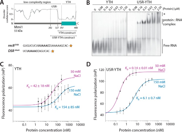
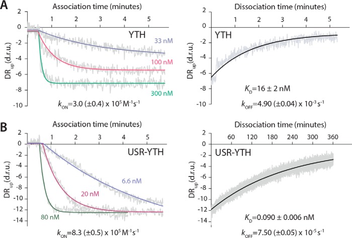
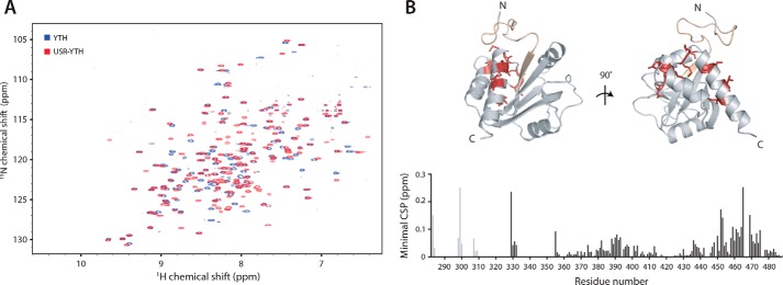
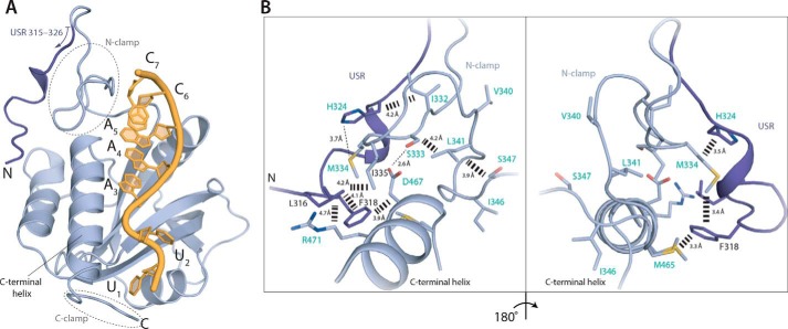
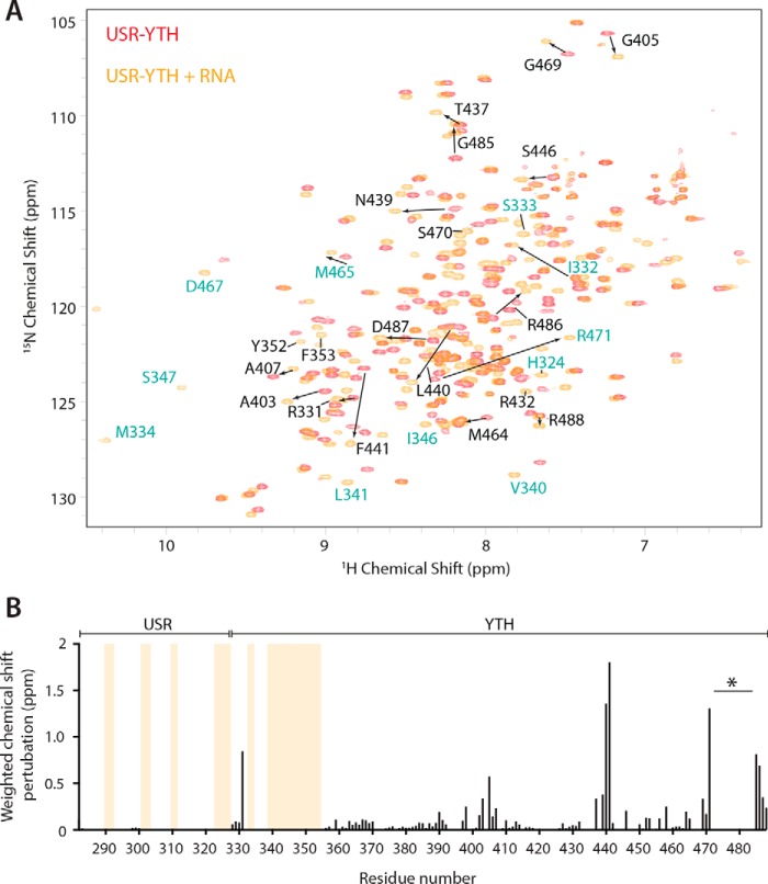
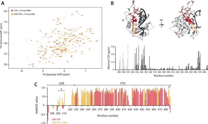
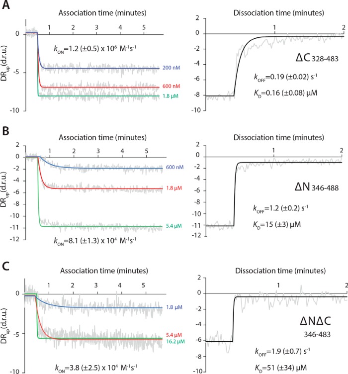
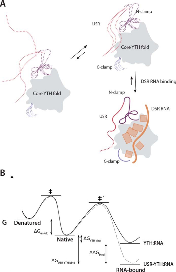
Mmi1 is an essential RNA-binding protein in the fission yeast Schizosaccharomyces pombe that eliminates meiotic transcripts during normal vegetative growth. Mmi1 contains a YTH domain that binds specific RNA sequences, targeting mRNAs for degradation. The YTH domain of Mmi1 uses a noncanonical RNA-binding surface that includes contacts outside the conserved fold. Here, we report that an N-terminal extension that is proximal to the YTH domain enhances RNA binding. Using X-ray crystallography, NMR, and biophysical methods, we show that this low-complexity region becomes more ordered upon RNA binding. This enhances the affinity of the interaction of the Mmi1 YTH domain with specific RNAs by reducing the dissociation rate of the Mmi1-RNA complex. We propose that the low-complexity region influences RNA binding indirectly by reducing dynamic motions of the RNA-binding groove and stabilizing a conformation of the YTH domain that binds to RNA with high affinity. Taken together, our work reveals how a low-complexity region proximal to a conserved folded domain can adopt an ordered structure to aid nucleic acid binding.

Keywords: RNA-binding protein; YT521-B homology; YTH domain; deadenylation; exosome specificity factor; intrinsically disordered protein; mRNA decay; meiosis; nuclear magnetic resonance (NMR); protein–nucleic acid interaction.


Conflict of interest statement

The authors declare that they have no conflicts of interest with the contents of this article

Figures

Figure 1.
Figure 1.
The USR increases the affinity of interaction of the Mmi1 YTH domain with RNA. A, top, schematic diagram of Mmi1 domain architecture, with the YTH and USR–YTH constructs indicated. The disorder prediction from DISOPRED3 is shown. Bottom, RNAs used for EMSAs (rec8DSR) and fluorescence polarization (DSRshort). The orange star represents the 3′-fluorescein label, and the UNAAAC motif is in bold. B, USR–YTH construct binds RNA more stably than the YTH domain alone. A fluorescently-labeled rec8DSR RNA containing the Mmi1 DSR motif was analyzed by EMSA after incubation with purified proteins at the indicated concentrations. Binding was analyzed by native PAGE. Free RNA, shifted protein–RNA complex, and higher-order supershifted complexes (asterisk) are indicated. C and D, fluorescence polarization assays of YTH (C) and USR–YTH (D) binding to DSRshort RNA (used at 0.1 nm). Calculated KD values are indicated, and error bars are the standard deviation of five biological replicates (each with three technical replicates).
Figure 2.
Figure 2.
The USR slows the off-rate of the YTH domain for RNA. Association and dissociation kinetics were calculated for the YTH (A) and USR–YTH (B) constructs. SwitchSENSE association binding curves at the indicated protein concentrations, plotted as dynamic response (DRup) in dynamic response units (d.r.u.) versus time are shown on the left. Dissociation curves, generated by flowing buffer across the chip surface saturated with YTH or USR–YTH protein, are shown on the right. Dynamic response (gray) is the integrated fluorescence intensity between 2 and 6 μs. Fitted exponential decay curves are shown with calculated rate constants and standard errors.
Figure 3.
Figure 3.
The USR alters the chemical environment of the C terminus of Mmi1. 2D NMR spectral analysis of YTH and USR–YTH constructs was performed. A, overlay of 1H-15N BEST-TROSY spectra of YTH and USR–YTH constructs. B, nearest neighbor chemical shift perturbation (CSP) maps of the assigned USR–YTH construct versus the unassigned YTH construct (bottom). Lighter gray peaks denote residues present in the USR–YTH construct but not the YTH construct. Regions that have large chemical shift differences (above 0.1 ppm; red) or are line broadened (yellow) are mapped onto the crystal structure of the Mmi1 YTH domain (top).
Figure 4.
Figure 4.
A network of interactions is formed between the USR, N-clamp, and core YTH domain. A, co-crystal structure of Mmi1 USR–YTH (residues 315–488 modeled) with DSR RNA. Cartoon representation shows protein in blue and RNA in yellow. The USR is shown in darker blue. B, conformation of the USR is stabilized by hydrophobic contacts with the core YTH domain and N-clamp. Residues labeled in turquoise either appear or show large chemical shift changes on RNA binding in NMR experiments (see Fig. 5).
Figure 5.
Figure 5.
The USR becomes more ordered on RNA binding. A, overlay of the 1H-15N BEST-TROSY spectra of USR–YTH Mmi1 in the absence (red) and presence (orange) of RNA. Selected assigned peaks that show large chemical shift perturbations (denoted with arrows) or appear on RNA binding are labeled. Residues labeled in turquoise are shown in Fig. 4B. B, plot showing the chemical shift perturbations per residue. Yellow bars indicate peaks that are only present in the RNA-bound form. The gap in the C-terminal region (asterisk) is due to line broadening in the presence of RNA. Other missing signals in the core YTH domain fold are due to incomplete back-exchange of the deuterated protein.
Figure 6.
Figure 6.
The USR influences the RNA-bound N- and C-clamps. 2D-NMR spectral analysis of YTH and USR–YTH constructs bound to a 19-mer DSR-containing RNA. A, overlay of 1H-15N BEST-TROSY spectra. B, nearest neighbor CSP maps (bottom). Lighter gray peaks denote residues present in the USR–YTH construct but not the YTH construct. Asterisks mark chemical shifts >0.3 ppm. Regions showing large chemical shift differences (above 0.1 ppm; red) or line broadening (yellow) are mapped onto the crystal structure of the Mmi1 YTH domain (top). C, plot of 1H-15N heteronuclear NOE values per residue in the absence (red) and presence (orange) of RNA. Positive values approaching 1 represent more ordered amide backbone regions. Peak absences represent residues that are not assigned. Many N-terminal residues that appear on RNA binding have hetNOE values that suggest they are ordered. The hash symbol marks some regions, assigned in both the absence and presence of RNA, with enhanced rigidity in the presence of RNA.
Figure 7.
Figure 7.
Low-complexity regions of Mmi1 are critical for high-affinity binding to DSR RNA. Binding kinetics for YTH-ΔC (A), YTH-ΔN (B), and YTH-ΔNΔC (C) constructs were determined using SwitchSENSE. Association (left panel) and dissociation (right panel) binding curves are shown, as dynamic response (DRup) versus time. Dynamic response is the integrated fluorescence intensity between 2 and 6 μs. Raw data are in gray, and fitted curves are in color or black.
Figure 8.
Figure 8.
Model for RNA binding by Mmi1. A, schematic diagram showing the contribution of different regions of Mmi1 to DSR RNA binding. The core, conserved YTH fold acts as a platform for RNA binding but is unable to bind RNA with high affinity alone. The USR, N-clamp, and C-clamps have no secondary structure and are dynamic in the absence of RNA (dashed lines). The USR and N-clamp likely exist in an equilibrium between multiple conformers, at least one of which has high affinity for RNA. On RNA binding, these regions become more ordered and contact or reinforce contacts with RNA. RNA backbone and bases are shown in orange. B, thermodynamic model of USR–YTH interaction with RNA. The presence of the USR (gray dashed line) does not affect the thermodynamic stability of the native protein compared with YTH alone (black line). Hence, as there is no change in the energy for unfolding (ΔGunfold) to the denatured state, the apparent melting temperatures are the same. The USR lowers the energy of the RNA-bound complex (ΔΔGbind) via changes in conformational dynamics and possibly a transient interaction with the RNA. Consequently, although the energy barrier for binding (native to ‡′) will be unaffected and similar association kinetics are observed with and without USR, the barrier for dissociation has increased, leading to significantly slower dissociation rates for USR–YTH/RNA compared with YTH/RNA.

References

    1. Harigaya, Y., Tanaka, H., Yamanaka, S., Tanaka, K., Watanabe, Y., Tsutsumi, C., Chikashige, Y., Hiraoka, Y., Yamashita, A., and Yamamoto, M. (2006) Selective elimination of messenger RNA prevents an incidence of untimely meiosis. Nature 442, 45–50 10.1038/nature04881 - DOI - PubMed
    1. Shichino, Y., Yamashita, A., and Yamamoto, M. (2014) Meiotic long non-coding meiRNA accumulates as a dot at its genetic locus facilitated by Mmi1 and plays as a decoy to lure Mmi1. Open Biol. 4, 140022–140022 10.1098/rsob.140022 - DOI - PMC - PubMed
    1. Yamashita, A., Shichino, Y., Tanaka, H., Hiriart, E., Touat-Todeschini, L., Vavasseur, A., Ding, D.-Q., Hiraoka, Y., Verdel, A., and Yamamoto, M. (2012) Hexanucleotide motifs mediate recruitment of the RNA elimination machinery to silent meiotic genes. Open Biol. 2, 120014–120014 10.1098/rsob.120014 - DOI - PMC - PubMed
    1. Cremona, N., Potter, K., and Wise, J. A. (2011) A meiotic gene regulatory cascade driven by alternative fates for newly synthesized transcripts. Mol. Biol. Cell. 22, 66–77 10.1091/mbc.E10-05-0448 - DOI - PMC - PubMed
    1. Mata, J., Lyne, R., Burns, G., and Bähler, J. (2002) The transcriptional program of meiosis and sporulation in fission yeast. Nat. Genet. 32, 143–147 10.1038/ng951 - DOI - PubMed

Publication types

MeSH terms