. 2018 May;592(10):1751-1760.
doi: 10.1002/1873-3468.13074. Epub 2018 May 15.

Chlamydial virulence factor TarP mimics talin to disrupt the talin-vinculin complex

Affiliations

Chlamydial virulence factor TarP mimics talin to disrupt the talin-vinculin complex

Austin J Whitewood et al. FEBS Lett. 2018 May.

Abstract

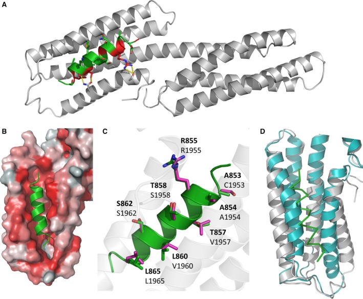
Vinculin is a central component of mechanosensitive adhesive complexes that form between cells and the extracellular matrix. A myriad of infectious agents mimic vinculin binding sites (VBS), enabling them to hijack the adhesion machinery and facilitate cellular entry. Here, we report the structural and biochemical characterisation of VBS from the chlamydial virulence factor TarP. Whilst the affinities of isolated VBS peptides from TarP and talin for vinculin are similar, their behaviour in larger fragments is markedly different. In talin, VBS are cryptic and require mechanical activation to bind vinculin, whereas the TarP VBS are located in disordered regions, and so are constitutively active. We demonstrate that the TarP VBS can uncouple talin:vinculin complexes, which may lead to adhesion destabilisation.

Keywords: adhesion; chlamydia; crystallography; molecular mimicry; talin; vinculin.


Figures

Figure 1
Figure 1
Biochemical characterisation of the TarP interaction with the vinculin head (Vd1). (A) Schematic of talin structure, with the location of the 11 talin VBS indicated (purple). (B) Schematic of TarP, indicating locations of VBS1 (green), VBS2 (orange), VBS3 (yellow), Actin binding site ABS (red) and LD‐motif (blue) at the C‐terminal. The disorder prediction trace generated using DISOPRED3 [44] is shown. (C) Multiple sequence alignment of vinculin binding sites, aligned using Clustal Omega. The consensus residues are highlighted in green. (D) Comparison of the Vd1:TarP and Vd1:talin interactions. Binding of fluorescein labelled talin VBS33, VBS36, TarP VBS (850–868)C and LD (655–680)C peptides to Vd1, measured using a fluorescence polarisation assay. Dissociation constants ± SEm) for the interactions are indicated in the legend. All measurements were performed in triplicate. ND, not determined.
Figure 2
Figure 2
Crystal structure of TarP in complex with Vd1. (A) Cartoon representation of the complex of Vd1 (grey) bound to TarP VBS (green); the consensus VBS residues are shown in red. (B) TarP VBS (green) docks into a hydrophobic groove on Vd1. Vd1 is represented as surface coloured by hydrophobicity: hydrophobic = red, hydrophilic = white. (C) TarP VBS peptide (green) aligned with talin VBS46 peptide (purple, PDB:1RKC 4) with Vd1‐interacting sidechains from both VBS shown as sticks and TarP residues (top bold) and corresponding vbs46 residues are shown. (D) VBS binding causes conformational change in the Vd1 domain. Comparison of apo Vd1 (cyan, PDB:1TR2 [45]) and TarP bound Vd1 (grey). The TarP peptide is shown as a ribbon (green).
Figure 3
Figure 3
TarP VBS disrupts the interaction between talin R10 and vinculin Vd1. Vd1 was incubated with talin R10 at 37 °C for 30 min then analysed on a gel filtration column (grey). The experiment was repeated with the addition of a stoichiometric amount of TarP VBS peptide (A) and then with talin VBS36 (B). All experiments were done in triplicate. 1% fluorescein‐labelled TarP VBS peptide was added to monitor TarP VBS elution at 494 nm which confirmed that TarP eluted bound to Vd1 (purple). (C) the relative ‘Weight Fraction’ percentage for talin:vinculin complex, talin, vinculin peaks in the absence and presence of both TarP VBS and VBS36 peptides. Data are means ± SEM; *P < 0.05 by T‐test. Both peptides reduced the R10‐Vd1 complex.
Figure 4
Figure 4
The TarP LD‐motif does not bind to FAK. (A) Multiple sequence alignment of known LD‐motifs and TarP, generated using Clustal Omega; the consensus binding residues are highlighted in blue. (B) Binding of fluorescein‐labelled TarP LD (655–680)C and Paxillin LD2 (141–153)C peptides to FAKFAT, measured using a fluorescence polarisation assay. Dissociation constants ± SEm) for the interactions are indicated in the legend. ND, not determined. (C) 1H,15N‐HSQC spectra of 130 μm 15N‐labelled FAKFAT in the absence (black) or presence of paxillin LD2 peptide (red; top panel) or TarP LD (green; bottom panel) at a ratio of 1 : 3.

References

    1. Calderwood DA, Campbell ID and Critchley DR (2013) Talins and kindlins: partners in integrin‐mediated adhesion. Nat Rev Mol Cell Biol 14, 503–517. - PMC - PubMed
    1. del Rio A, Perez‐Jimenez R, Liu R, Roca‐Cusachs P, Fernandez JM and Sheetz MP (2009) Stretching single talin rod molecules activates vinculin binding. Science (80‐.) 323, 638–641. - PMC - PubMed
    1. Yao M, Goult BT, Klapholz B, Hu X, Toseland CP, Guo Y, Cong P, Sheetz MP and Yan J (2016) The mechanical response of talin. Nat Commun 7, 11966. - PMC - PubMed
    1. Izard T, Evans G, Borgon RA, Rush CL, Bricogne G and Bois PR (2004) Vinculin activation by talin through helical bundle conversion. Nature 427, 171–175. - PubMed
    1. Carisey A, Tsang R, Greiner AM, Nijenhuis N, Heath N, Nazgiewicz A, Kemkemer R, Derby B, Spatz J and Ballestrem C (2013) Vinculin regulates the recruitment and release of core focal adhesion proteins in a force‐dependent manner. Curr Biol 23, 271–281. - PMC - PubMed

Publication types

LinkOut - more resources