. 2023 Sep 6;6(11):e202201784.
doi: 10.26508/lsa.202201784. Print 2023 Nov.

Structure shows that the BIR2 domain of E3 ligase XIAP binds across the RIPK2 kinase dimer interface

Affiliations

Structure shows that the BIR2 domain of E3 ligase XIAP binds across the RIPK2 kinase dimer interface

Mathilde Lethier et al. Life Sci Alliance. .

Abstract

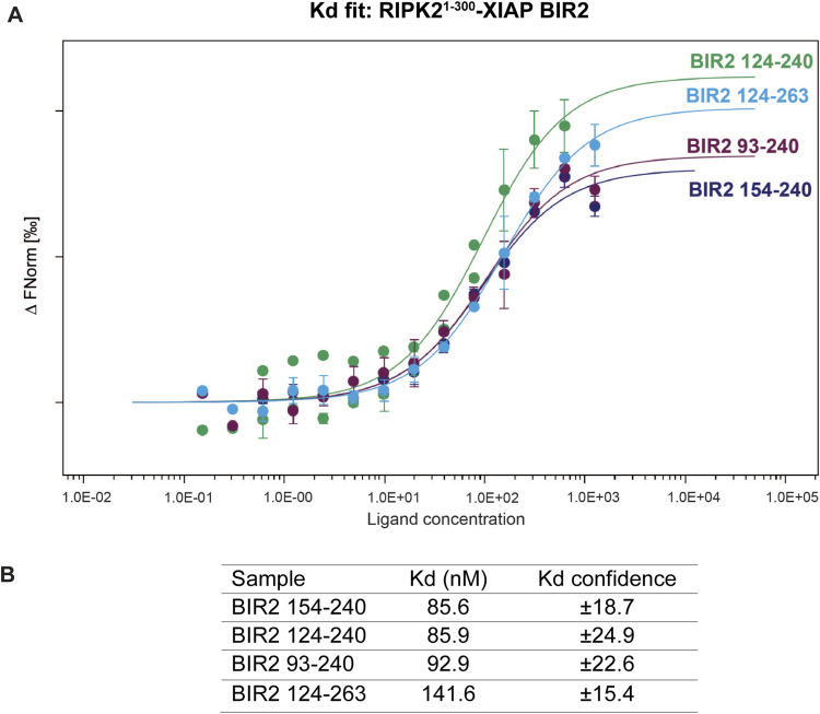
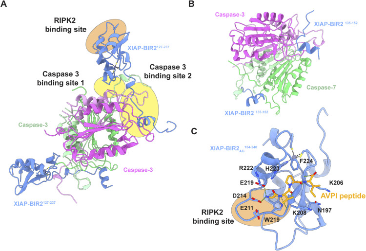
RIPK2 is an essential adaptor for NOD signalling and its kinase domain is a drug target for NOD-related diseases, such as inflammatory bowel disease. However, recent work indicates that the phosphorylation activity of RIPK2 is dispensable for signalling and that inhibitors of both RIPK2 activity and RIPK2 ubiquitination prevent the essential interaction between RIPK2 and the BIR2 domain of XIAP, the key RIPK2 ubiquitin E3 ligase. Moreover, XIAP BIR2 antagonists also block this interaction. To reveal the molecular mechanisms involved, we combined native mass spectrometry, NMR, and cryo-electron microscopy to determine the structure of the RIPK2 kinase BIR2 domain complex and validated the interface with in cellulo assays. The structure shows that BIR2 binds across the RIPK2 kinase antiparallel dimer and provides an explanation for both inhibitory mechanisms. It also highlights why phosphorylation of the kinase activation loop is dispensable for signalling while revealing the structural role of RIPK2-K209 residue in the RIPK2-XIAP BIR2 interaction. Our results clarify the features of the RIPK2 conformation essential for its role as a scaffold protein for ubiquitination.


Conflict of interest statement

The authors declare that they have no conflict of interest.

Figures

Figure 1.
Figure 1.. In vitro reconstitution and stoichiometry of the RIPK2–XIAP BIR2 complex.
(A) Overview of the XIAP BIR2 constructs used in this article. (B) SEC profile and SDS–PAGE gel of RIPK21−317–XIAP BIR2154–240 complex. Uncropped SDS–PAGE gel is reported in Fig S1B. (C) Native mass spectrometry results for short BIR2 (top spectra) and long BIR2 (bottom spectra) constructs. The main stoichiometry ratios are 2:1 and 2:2 (RIPK2:XIAP BIR2), where two molecules of RIPK2 are required for the interaction with XIAP BIR2. The stoichiometry becomes 2:2 in the case of RIPK2- XIAP BIR2AG124240.
Figure S1.
Figure S1.. In vitro reconstitution of the RIPK2–XIAP BIR2 complex.
(A, B, C, D) Size-exclusion chromatography profile of the 4 in vitro-reconstituted RIPK2–XIAP BIR2 complexes together with Comassie-stained SDS–PAGE analysis of eluted fractions: (A) RIPK21−300-XIAP BIR2124−240, (B) RIPK21−317-XIAP BIR2154−240, (C) RIPK21−317-XIAP BIR2AG124240, (D) RIPK21−317-XIAP BIR2AG154240. The size-exclusion chromatography profile of RIPK21−317-XIAP BIR2154−240, already shown in Fig 1B, is reported here for completeness.
Figure S2.
Figure S2.. RIPK2–XIAP BIR2 dissociation constants measured by MST.
(A) Dose-response curves of wild-type XIAP BIR2 constructs toward RIPK21−300. All experiments have been done in triplicates. Standard deviations are represented for each point in bars. (B) The table reports the Kd obtained for each sample. All the dose-response curves were fitted to a one-site–binding model to obtain Kd value.
Figure 2.
Figure 2.. NMR characterization of the XIAP BIR2 interaction with RIPK2 kinase in solution.
(A) Overlap of 1H-15N BEST-TROSY correlation spectra (25°C) recorded at 850 MHz 1H frequency on samples of 15N-labeled XIAP BIR2 (black) and a 1:1 complex of 15N-labeled XIAP BIR2 and unlabeled RIPK21−317 (red). BIR2 residues that remain visible in the complex are annotated by their amino acid type (one-letter code) and residue number. No or only small chemical shift changes are detected for these residues between the free BIR2 protein and the complex, indicating that these protein segments are not involved in the interaction. (B) Surface representation of the XIAP BIR2 structure (PDB ID: 1C9Q, Sun et al, 1999). The N- and C-terminal protein segments that are not involved in the interaction with RIPK2 are highlighted in red. (C) {1H}-15N heteronuclear NOE (HETNOE) ratios measured for XIAP BIR2 at 25°C and 850 MHz 1H frequency. The protein segments that remain visible in the NMR correlation spectrum of (A) upon interaction with RIPK2 are highlighted by red bars. These N- and C-terminal segments show reduced HETNOE ratios (≤0.6), indicative of significant fast (sub-ns) time scale backbone mobility, whereas for the central part, an average HETNOE of 0.8 is measured, in agreement with a globular protein domain. The flexibility of the N- and C-terminal segments is preserved in the complex, which makes them NMR-observable despite their high molecular weight.
Figure S3.
Figure S3.. Translational diffusion properties of XIAP BIR2 (black) and the RIPK21−317–XIAP BIR2AG124–240 complex (red) measured by 1D 1H DOSY at 25°C and 850 MHz 1H frequency.
Exponential fitting of these decay curve results in diffusion coefficients of 13.6 10−7 m2s−1 for XIAP BIR2 and 7.6 10−7 m2 s−1 for RIPK2:XIAP BIR2. The translational diffusion coefficient provides a measure of the average particle size in solution. Upon interaction with RIPK2, the apparent molecular size is increased by a factor of about 5.7, in good agreement with a 1:2 RIPK2:BIR2 stoichiometry.
Figure S4.
Figure S4.. RIPK21−317–XIAP BIR2AG154–240 complex purification for cryo-EM.
(A) RIPK2 and XIAP BIR2 constructs used for cryo-EM structure determination. (B) Zoom-in on the RIPK21−317–XIAP BIR2AG154240 SEC profile of Fig S1B; collected fractions are shown in magenta; fractions 3–6 have been used for cryo-EM specimen preparation. (C) Exemplary micrograph collected during specimen screening (Glacios TEM equipped with Falcon3, counting mode, −1 μm defocus, pixel size 0.94 px/Å, 40 e−/A2 total dose). (D) Exemplary micrographs collected at Krios in either tilted or untilted mode (EF, energy filter). Dose and pixel size are reported in the main text and in Table S2.
Figure S5.
Figure S5.. Cryo-EM processing workflow.
The scheme summarizes the cryo-EM processing to obtain the final map, as described in the Materials and Methods section. The unsharpened maps, the FSC threshold, and the angular sampling schemes show how the map improved along the processing, without losing orientations and gaining in the density at the interaction between RIPK2 kinase (Kinase_A in light grey, Kinase_B in dark grey) and XIAP BIR2 (in light blue).
Figure S6.
Figure S6.. Local resolution of the RIPK21−317-XIAP BIR2154−240 map.
(A) Directional FSC plot calculated in CryoSPARC. (B) Masked FSC half-maps and masked FSC map-model computed in Phenix (Afonine et al, 2018). (C) Cryo-EM density sharpened at B-factor −100 Å2 of the RIPK21−317-XIAP BIR2154−240 complex colored according to local resolution. Local resolution has been estimated in CryoSPARC.
Figure 3.
Figure 3.. Structure of the RIPK21−317–XIAP BIR2154−240 complex.
(A) Ribbon representation of the RIPK21−317-XIAP BIR2154−240 structure. XIAP BIR2 is colored in light blue, kinases molecules in light and dark grey (Kinase_A, Kinase_B), αC-helix in pink (residues 52–72), Gly-rich loop in yellow, activation loop in green, and K209 loop in magenta (as described in Pellegrini et al, 2017). (B, C, D, E, F) Assignment according to the cryo-EM density features of (B) Kinase_A αE-helix residues (120–137), (C) Kinase_B αE-helix residues (120–137), (D) Kinase_A and Kinase_B αL-helices and their interaction (residues 299–312), (E) XIAP BIR2 interaction with Kinase_A, (F) XIAP BIR2 interaction with Kinase_B.
Figure S7.
Figure S7.. Fitting of the RIPK21−317–XIAP BIR2154−240 structure in the density map.
(A, B) Fitting of (A) Kinase_A and (B) XIAP BIR2 domain in the cryo-EM density sharpened at B-factor−100 Å2. (C, D) Cryo-EM density corresponding to the non-hydrolysable ATP analogue ACP in (C) Kinase_A and (D) Kinase_B. The fitting has been obtained by fitting in chimeraX the active structure of RIPK2 kinase (PDB ID: 5NG0) (Pellegrini et al, 2017). Chains and smaller elements are colored as in Fig 3A.
Figure S8.
Figure S8.. Flexible RIPK2 N-terminus improves structure fitting in the EM density.
(A, B) Comparison of fitting in the cryo-EM density between RIPK2 dimer belonging to the RIPK21−317–XIAP BIR2154−240 structure (A) and active RIPK21−300 structure (PDB ID: 5NG0) (Pellegrini et al, 2017) (B). Structures have been aligned on Kinase_B molecule of RIPK21−317-XIAP BIR2154−240. (A) Left: ribbon representation of the RIPK21−317-XIAP BIR2154−240 structure, with view on kinase αC-helixes and N-termini. Chains and smaller elements are colored as in Fig 3A. Middle: fitting of the αC-helixes (residues 57–72), the activation loops (residues 169–171), and kinase N-termini (residues 8–10) in the cryo-EM density. Right: fitting in the density of the same protein elements belonging to the crystallographic RIPK21−300 structure (PDB ID: 5NG0). In 5NG0, the N-terminus comprises residues 6–10. (B) Left: ribbon representation of the RIPK21−317–XIAP BIR2154−240 structure, rotated 180°. Middle: fitting of residues 90–105 from Kinase_ A and 73–82 from Kinase_B in the cryo-EM density. Right: fitting in the density of the same protein elements belonging to the crystallographic RIPK21−300 structure (PDB ID: 5NG0). (C) Left: alphafold2 prediction of the RIPK21−317-XIAP BIR2154−240 structure, colored according to prediction confidence (blue = 100, yellow = 70, orange = 50, red = 0). Middle: zoom-in on the αC-helixes and N-termini. Right: predicted aligned error diagram of alphafold2 prediction.
Figure 4.
Figure 4.. Validation of the interaction interfaces between RIPK2 and XIAP BIR2.
(A) Schematic representation of the RIPK2 and XIAP constructs used for the expression of wt and mutant proteins in HEKT293 cells. (B, C) Results of expression and pull down (IP, immunoprecipitation) on HA-RIPK2 and MYC-XIAP proteins from HEKT293 cells. Lines corresponding to IPs against HA and β-actin are in blue, whereas lanes corresponding to IP against MYC re in green. IPs have been repeated twice. (D, E) Reconstitution of RIPK2−/− iBMDMs with doxycycline-inducible forms of RIPK2. (D) Cells were transduced with doxycylcine-inducible lentiviral vectors expressing WT or N137L RIPK2. After selection, clones were pooled. They were then left untreated or treated overnight with 500 ng/ml doxycycline. Western blotting was performed and showed equivalently inducible levels of RIPK2 WT and RIPK2 N137L. (E) The cells generated in Panel A were then left untreated or were treated with 500 ng/ml doxycycline overnight. After this treatment, cells were either not exposed or exposed to 10 mg/ml MDP for 4 h qRT–PCR was then performed using the NOD2-inducible IRG1 and CXCL10 genes. Biological triplicates, each with two technical replicates, are shown. Source data are available for this figure.
Figure S9.
Figure S9.. Binding of RIPK2 mutants to XIAP BIR2.
(A, B, C, D) SEC profile of (A) RIPK21−317-XIAP BIR2154–240, (B) RIPK21−317 K209A-XIAP BIR2, (C) RIPK21−317 K209R-XIAP BIR2154−240, and (D) RIPK21−317 S282L-XIAP BIR2154−240 with Comassie-stained SDS–PAGE analysis of eluted fractions. Source data are available for this figure.
Figure S10.
Figure S10.. Binding of the XIAP BIR2 domain to caspase 3, caspase 7, and the AVPI peptide.
(A) Ribbon representation of the XIAP BIR2–caspase 3 complex (PDB ID: 1I3O) (Riedl et al, 2001) The BIR2 domain of XIAP (colored in blue) binds backward to the catalytic subunit of caspase 3 compared with RIPK2 binding (see the orange and yellow ovals). Using BIR2 residues N226, F228, R233 (caspase 3-binding site 1) and the linker region (caspase 3-binding site 2), XIAP interacts with both catalytic subunits of caspase 3 (colored in pink and green). Specifically, each XIAP BIR2 molecule (residues 127–237) interacts with both the large and small subunits of one caspase molecule (colored in dark and light pink, respectively) and with a short stretch of the adjacent caspase molecule (colored in dark and light green). (B) Ribbon representation of the XIAP BIR2–caspase 7 complex (PDB ID: 1I51) (Chai et al, 2001). XIAP BIR2 interacts with both large and small domains of the catalytic subunit of caspase 7 (colored in dark pink/green and light pink/green, respectively) using the linker region (residues 135–152, colored in blue). (C) Ribbon representation of XIAP BIR2 domain bound to AVPI peptide (PDB ID: 4J46) (Lukacs et al, 2013). The XIAP BIR2 residues involved in the binding to the peptide are highlighted in stick (IBM grove). As shown by the orange oval, the binding to RIPK2 overlaps with the binding to the SMAC peptide. Zn is represented in the grey sphere. Metal ion bonds are represented in black dashed line, whereas H-bonds are represented in green dashed line.
Figure S11.
Figure S11.. Conformation of DFG motif and activation loop in ligand-bound RIPK2 structures.
(A, B, C, D) Ribbon representation of (A) RIPK2317–XIAP BIR2154−240 structure (this study) and RIPK2 kinase domain in complex with (B) CSLP18 (PDB ID: 6FU5) (Hrdinka et al, 2018), (C) ponatinib (PDB ID: 4C8B) (Canning et al, 2015), (D) GSK583 (PDB ID: 5J7B) (Haile et al, 2016). Each figure shows the conformation of the DFG motif (residues 164–166, in stick) and the activation loop if present. The αC-helix is colored in pink, the Gly-rich loop in yellow, the activation loop in green, and the K209 loop in magenta, as in Fig 3.
Figure S12.
Figure S12.. Alphafold2 predictions of cIAP1 BIR2, cIAP2 BIR2, RIPK2-cIAP1 BIR2, and RIPK2-cIAP2 BIR2 structures.
(A) Structure-based alignment of cIAPs BIR2 domains, within the assigned Uniprot code. The alignment was computed using Clustal Omega (Madeira et al, 2022) and the output was used as input for ESPript 3.0, to generate the figure (Robert & Gouet, 2014). The secondary structure assignment of the XIAP BIR2 crystallographic structure is shown on top (PDB ID: 4J3Y) (Lukacs et al, 2013). Residues with consensus >70 are highlighted in red. The high similarity with the XIAP loop 200–214 is highlighted in blue (NWExxDx motif). (B) Ribbon representation of the crystallographic XIAP BIR2 structure and the Alphafold2-predicted structure of cIAP1 BIR2 and cIAP2 BIR2 domains (from Alphafold2 database). Side chains of the NWExxDx motif are shown. (C) Ribbon representation of the RIPK21−317-XIAP BIR2154−240 structure (this article) and the Alphafold2-predicted structures of RIPK21−317–cIAP1 BIR2 and RIPK21−317–cIAP2 BIR2 complexes. Alphafold2-predicted models are coloured according to prediction confidence (blue = 100, yellow = 70, orange = 50, red = 0). The predicted aligned error diagram is shown for the structures computed by us.

References

    1. Abbott DW, Wilkins A, Asara JM, Cantley LC (2004) The Crohn[R8S2Q1M7]s disease protein, NOD2, requires RIP2 in order to induce ubiquitinylation of a novel site on NEMO. Curr Biol 14: 2217–2227. 10.1016/J.CUB.2004.12.032 - DOI - PubMed
    1. Afonine PV, Klaholz BP, Moriarty NW, Poon BK, Sobolev OV, Terwilliger TC, Adams PD, Urzhumtsev A (2018) New tools for the analysis and validation of cryo-EM maps and atomic models. Acta Crystallogr D Struct Biol 74: 814–840. 10.1107/S2059798318009324 - DOI - PMC - PubMed
    1. Amar J, Chabo C, Waget A, Klopp P, Vachoux C, Bermúdez-Humarán LG, Smirnova N, Bergé M, Sulpice T, Lahtinen S, et al. (2011) Intestinal mucosal adherence and translocation of commensal bacteria at the early onset of type 2 diabetes: Molecular mechanisms and probiotic treatment. EMBO Mol Med 3: 559–572. 10.1002/emmm.201100159 - DOI - PMC - PubMed
    1. Ananthakrishnan AN (2015) Epidemiology and risk factors for IBD. Nat Rev Gastroenterol Hepatol 12: 205–217. 10.1038/nrgastro.2015.34 - DOI - PubMed
    1. Atreya I, Atreya R, Neurath MF (2008) NF-kappaB in inflammatory bowel disease. J Intern Med 263: 591–596. 10.1111/j.1365-2796.2008.01953.x - DOI - PubMed

Publication types

Substances

LinkOut - more resources