. 2019 Nov 8;294(45):17143-17154.
doi: 10.1074/jbc.RA119.010842. Epub 2019 Sep 23.

Structural basis for the specific cleavage of core-fucosylated N-glycans by endo-β- N-acetylglucosaminidase from the fungus Cordyceps militaris

Affiliations

Structural basis for the specific cleavage of core-fucosylated N-glycans by endo-β- N-acetylglucosaminidase from the fungus Cordyceps militaris

Haruka Seki et al. J Biol Chem. .

Abstract

N-Linked glycans play important roles in various cellular and immunological events. Endo-β-N-acetylglucosaminidase (ENGase) can release or transglycosylate N-glycans and is a promising tool for the chemoenzymatic synthesis of glycoproteins with homogeneously modified glycans. The ability of ENGases to act on core-fucosylated glycans is a key factor determining their therapeutic utility because mammalian N-glycans are frequently α-1,6-fucosylated. Although the biochemistries and structures of various ENGases have been studied extensively, the structural basis for the recognition of the core fucose and the asparagine-linked GlcNAc is unclear. Herein, we determined the crystal structures of a core fucose-specific ENGase from the caterpillar fungus Cordyceps militaris (Endo-CoM), which belongs to glycoside hydrolase family 18. Structures complexed with fucose-containing ligands were determined at 1.75-2.35 Å resolutions. The fucose moiety linked to GlcNAc is extensively recognized by protein residues in a round-shaped pocket, whereas the asparagine moiety linked to the GlcNAc is exposed to the solvent. The N-glycan-binding cleft of Endo-CoM is Y-shaped, and several lysine and arginine residues are present at its terminal regions. These structural features were consistent with the activity of Endo-CoM on fucose-containing glycans on rituximab (IgG) and its preference for a sialobiantennary substrate. Comparisons with other ENGases provided structural insights into their core fucose tolerance and specificity. In particular, Endo-F3, a known core fucose-specific ENGase, has a similar fucose-binding pocket, but the surrounding residues are not shared with Endo-CoM. Our study provides a foothold for protein engineering to develop enzymatic tools for the preparation of more effective therapeutic antibodies.

Keywords: Cordyceps militaris; GH18; N-linked glycosylation; X-ray crystallography; core-fucosylated N-glycan; endo-β-N-acetylglucosaminidase (ENGase); glycoprotein; glycoside hydrolase; monoclonal antibody; substrate-assisted mechanism.


Conflict of interest statement

The authors declare that they have no conflicts of interest with the contents of this article

Figures
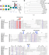
Figure 1.
Figure 1.
Reaction and diversity of ENGases. A, hydrolytic cleavage site of an N-glycan by Endo-CoM. The best substrate for Endo-CoM (core-fucosylated sialobiantennary) is shown by the Symbol Nomenclature for Glycans (65). The Fuc–GlcNAc–Asn moiety bound to the Endo-CoM structure and the octasaccharide of biantennary complex glycan observed in other ENGase structures are boxed by dashed lines. Subsites of ENGases are indicated by blue characters. B, phylogenetic tree of GH18 ENGases. The fucose-specific ENGases and structure-known ones are selected. Bar, 5% sequence divergence. C, partial amino acid sequence alignment of GH18 ENGases. The catalytic residues and key amino acid residues for the Fuc–GlcNAc recognition of Endo-CoM are indicated above the sequences with red and blue arrows, respectively.
Figure 2.
Figure 2.
Crystal structure of Endo-CoM. A, overall structure of D154N/E156Q mutant (ribbon model with rainbow colors) complexed with Fuc–GlcNAc–Asn (white sticks). The side chains of the mutated catalytic residues (Asn-154 and Gln-156) are shown as magenta sticks. A disulfide bond (Cys-24–Cys-76) connecting the additional N-terminal α-helix (blue) is shown as a stick with the sulfur atoms in yellow. B–D, electron density maps of the complex structures. mFoFc omit maps for Fuc (B, 3.0σ), Fuc–GlcNAc (C, 3.0σ), and Fuc–GlcNAc–Asn (D, 1.5σ) are shown. D, Fuc–GlcNAc–FmocAsn was used for the crystal soaking but the Fmoc moiety was disordered. E, comparison of the apo and complex structures at the active site. The apo structure (gray) and the complex structures with Fuc (cyan), Fuc–GlcNAc (yellow), and Fuc–GlcNAc–Asn (green) near subsites +1 and +1′ are superimposed. The ligands and hydrogen bonds are shown with thick sticks and dashed lines (magenta), respectively.
Figure 3.
Figure 3.
Stereoviews of the active site of Endo-CoM complexed with Fuc–GlcNAc–Asn. The protein main chain is shown as a gray ribbon model. The side chains near the ligand, Fuc, and GlcNAc–Asn moieties of the ligand are shown as sticks colored in green, cyan, and yellow, respectively. A, biantennary complex glycan bound to Endo-F3 (PDB ID: 1EOM) is shown as thin blue sticks. B, chitin pentamer bound to ChiB (1E6N) is shown as blue sticks. The N,N′-diacetylchitobiose unit bound at subsites −1 and +1 is highlighted with thick sticks. The side chains of the catalytic residues of Endo-F3 and ChiB are also shown as blue sticks.
Figure 4.
Figure 4.
Comparison of the fucose-binding site of Endo-CoM (A) with a potential subsite +1′ of Endo-F3 (B) and Endo-S (C). In B (1EOM) and C (6EN3), bound biantennary complex glycan is shown as thin blue sticks, and the Fuc–GlcNAc–Asn ligand of Endo-CoM is overlaid.
Figure 5.
Figure 5.
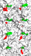
Molecular surfaces of Endo-CoM (A), Endo-F3 (B, 1EOM), Endo-S (C, 6EN3), Endo-S2 (D, 6MDS), Endo-H (E, 1EDT), Endo-F1 (F, 2EBH), BT1044 (G, 6Q64), and Endo-T (H, 4AC1) at the fucose-binding site. The catalytic residues (Asp-154 and Glu-156 in Endo-CoM) and the conserved Tyr residue (Tyr-216 in Endo-CoM), which were used for superimposition of the protein structures, are colored in red and green, respectively. Biantennary complex glycan bound to Endo-F3, Endo-S, and Endo-S2 are shown as thin blue sticks, and the Fuc–GlcNAc–Asn ligand of Endo-CoM is overlaid on the structures of B–H. Residues that potentially form a steric crash with the fucose are indicated by purple characters.
Figure 6.
Figure 6.
Glycan-binding clefts of Endo-CoM (A and B), Endo-F3 (C, 1EOM), Endo-S (D, 6EN3), and Endo-S2 (E). The color code for the loops is according to Ref. . The biantennary complex glycan bound to the three ENGases (blue sticks) is also schematically shown in a blue box. C, potential binding area for the Gal–GlcNAc-branch of triantennary complex-type glycans is indicated by an orange circle. E, molecular surface of Endo-S2 complexed with the biantennary complex glycan (6MDS) is overlaid with the high-mannose glycan (salmon sticks) bound to the same protein (6MDV). The high-mannose glycan is also schematically shown in a salmon box. A pocket for accommodating the α1,3-branched antenna is indicated by a red circle.

References

    1. Bieberich E. (2014) in Glycobiology of the Nervous System (Yu R. K., and Schengrund C.-L., eds) pp. 47–70, Springer, New York: 10.1007/978-1-4939-1154-7_3 - DOI
    1. Maverakis E., Kim K., Shimoda M., Gershwin M. E., Patel F., Wilken R., Raychaudhuri S., Ruhaak L. R., and Lebrilla C. B. (2015) Glycans in the immune system and the altered glycan theory of autoimmunity: a critical review. J. Autoimmun. 57, 1–13 10.1016/j.jaut.2014.12.002 - DOI - PMC - PubMed
    1. Stanley P., Taniguchi N., and Aebi M. (2017) Essentials of Glycobiology, 3rd Ed, Chapter 9, Cold Spring Harbor Laboratory Press, Cold Spring Harbor, NY: 10.1101/glycobiology.3e.009 - DOI
    1. Pučić M., Knežević A., Vidič J., Adamczyk B., Novokmet M., Polašek O., Gornik O., Šupraha Goreta S., Wormald M. R., Redžic I., Campbell H., Wright A., Hastie N. D., Wilson J. F., Rudan I., et al. (2011) High throughput isolation and glycosylation analysis of IgG-variability and heritability of the IgG glycome in three isolated human populations. Mol. Cell. Proteomics 10, M111.010090 10.1074/mcp.M111.010090 - DOI - PMC - PubMed
    1. Takahashi M., Kuroki Y., Ohtsubo K., and Taniguchi N. (2009) Core fucose and bisecting GlcNAc, the direct modifiers of the N-glycan core: their functions and target proteins. Carbohydr. Res. 344, 1387–1390 10.1016/j.carres.2009.04.031 - DOI - PubMed

Publication types