. 2020 Dec 25;295(52):18579-18588.
doi: 10.1074/jbc.RA120.015303. Epub 2020 Oct 29.

High affinity binding of SARS-CoV-2 spike protein enhances ACE2 carboxypeptidase activity

Affiliations

High affinity binding of SARS-CoV-2 spike protein enhances ACE2 carboxypeptidase activity

Jinghua Lu et al. J Biol Chem. .

Abstract

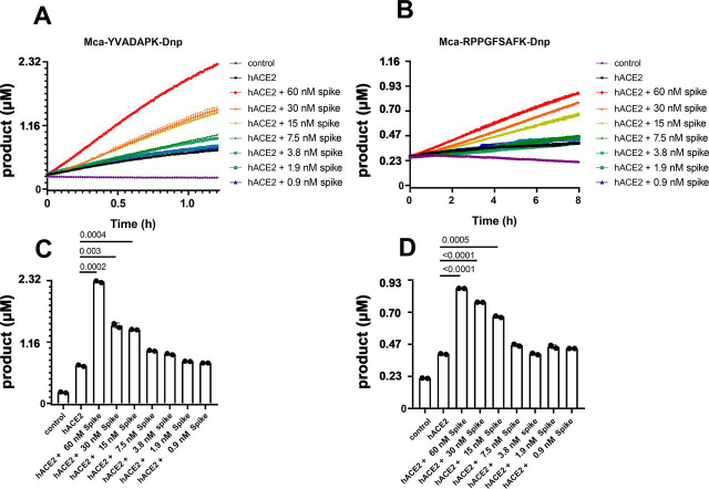
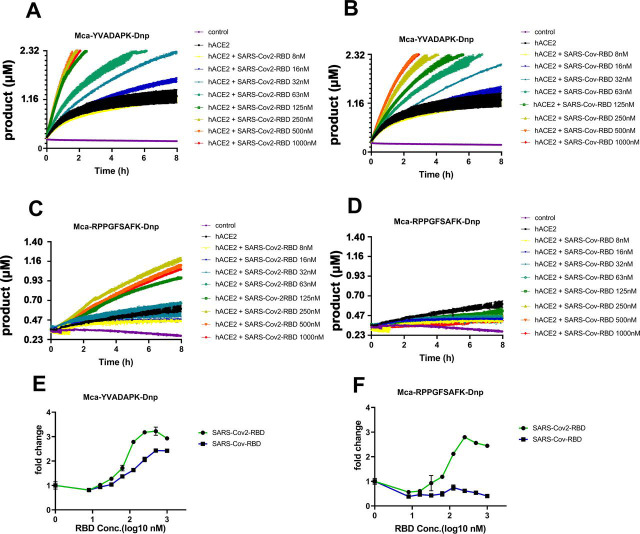
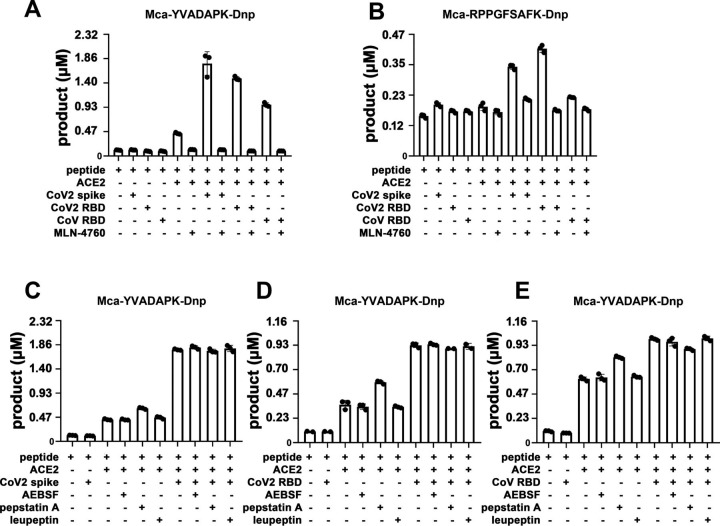
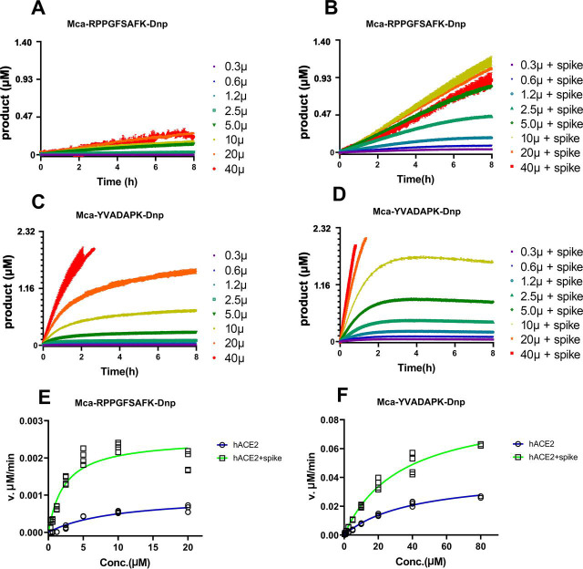
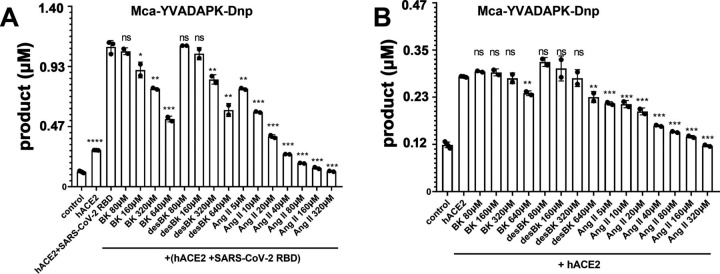
The novel severe acute respiratory syndrome coronavirus (SARS-CoV-2) has emerged to a pandemic and caused global public health crisis. Human angiotensin-converting enzyme 2(ACE2) was identified as the entry receptor for SARS-CoV-2. As a carboxypeptidase, ACE2 cleaves many biological substrates besides angiotensin II to control vasodilatation and vascular permeability. Given the nanomolar high affinity between ACE2 and SARS-CoV-2 spike protein, we investigated how this interaction would affect the enzymatic activity of ACE2. Surprisingly, SARS-CoV-2 trimeric spike protein increased ACE2 proteolytic activity ∼3-10 fold against model peptide substrates, such as caspase-1 substrate and Bradykinin-analog. The enhancement in ACE2 enzymatic function was mediated by the binding of SARS-CoV-2 spike RBD domain. These results highlighted the potential for SARS-CoV-2 infection to enhance ACE2 activity, which may be relevant to the cardiovascular symptoms associated with COVID-19.

Keywords: SARS-CoV-2 spike protein; angiotensin converting enzyme 2; carboxypeptidase; enzymatic activity; fluorescence resonance energy transfer (FRET); pathogenesis; renin angiotensin system; viral protein.


Conflict of interest statement

Conflict of interest—The authors declare that they have no conflicts of interest with the contents f this article.

Figures

Figure 1
Figure 1
SARS-CoV-2 spike protein enhanced ACE2 cleavage of fluorogenic caspase-1 substrate and bradykinin analog in a concentration dependent manner.A and B, Kinetic reading of the hydrolysis of Mca-YVADAPK-Dnp(20 μm) and Mca-RPPGFSAFK-Dnp (20 μm) in the presence of SARS-CoV-2 spike protein at indicated final concentrations in the enzymatic assays. The maximum reading is limited to 100000 RFU on Synergy H1 plate reader. RFU was converted to product concentrations with 1 μm equal to 42942 RFU based on the calibration standard. C and D, Comparison of the cleavage of Mca-YVADAPK-Dnp and Mca-RPPGFSAFK-Dnp at 1.5h and 8 h, respectively. Individual p-values for pairwise students' t test were displayed between each pair. The mean and standard deviation of each reaction were shown as columns with each RFU reading repetition displayed as a black dot.
Figure 2
Figure 2
SARS-CoV-2 RBD but not SARS-CoV RBD enabled ACE2 to cleave bradykinin analog.A and B, hydrolysis of Mca-YVADAPK-Dnp(20 μm) in the presence of SARS-CoV-2 RBD and SARS-CoV RBD proteins over time at indicated concentrations. C and D, hydrolysis of Mca-RPPGFSAFK-Dnp (20 μm) in the presence of SARS-CoV-2 RBD and SARS-CoV RBD proteins at indicated concentrations. Data are shown as relative fluorescence units (RFU) where the maximum reading is 100000 RFU on Synergy H1 plate reader. E, Fold change of RFU during Mca-YVADAPK-Dnp cleavage in the presence of SARS-CoV-2 RBD or SARS-CoV RBD at the time point of 1.5h because of instrument overflow. F, Fold change of RFU during Mca-RPPGFSAFK-Dnp cleavage in the presence of SARS-CoV-2 RBD or SARS-CoV RBD at time point of 8h. All RFU readings at different concentrations of RBD proteins were normalized to that of ACE2 cleavage without RBD proteins correspondingly.
Figure 3
Figure 3
A and B, ACE2 specific inhibitor MLN-4760 (10 μm) completely blocked ACE2 activity and abolished the enhancement mediated by SARS-CoV-2 spike and RBD protein binding.CE, various protease inhibitors at 40 μm (AEBSF, pepstatin A, leupeptin) had no effects on ACE2 activity.
Figure 4
Figure 4
Measurement of kinetic constants for hydrolysis of Mca-RPPGFSAFK-Dnp and Mca-YVADAPK-Dnp in the absence or presence of SARS-CoV-2 spike protein (14 µg/ml).A and B, ACE2 hydrolysis of Mca-RPPGFSAFK-Dnp at different concentrations in the absence (A) and presence (B) of SARS-CoV-2 spike protein. C and D, ACE2 hydrolysis of Mca-YVADAPK-Dnp at different concentrations in the absence (C) and presence (D) of SARS-CoV-2 spike protein. Background RFU readings were subtracted. E and F, Michaelis plots for ACE2 hydrolysis of Mca-RPPGFSAFK-Dnp and Mca-YVADAPK-Dnp in the absence (blue) or presence (green)of SARS-CoV-2 spike protein. The initial velocity conditions were limited to 30 min for Mca-YVADAPK-Dnp and 60 min for Mca-RPPGFSAFK-Dnp because of different cleavage rate. All determinations were repeated with duplicates.
Figure 5
Figure 5
Competition of Bradykinin(BK), des-Arg9-BK, and Ang II peptide with fluorogenic Mca-YVADAPK-Dnp (10 μm) for ACE2 hydrolysis.A, Competitive substrates inhibition in the presence of SARS-CoV-2 RBD protein at time 1hof fluorogenic Mca-YVADAPK-Dnp cleavage. The pairwise p-value statistics were calculated between (hACE2 + SARS-CoV-2 RBD) and its addition of corresponding concentrations of competitive peptides. B, Competitive substrates inhibition in the absence of SARS-CoV-2 RBD protein at time 1 h of fluorogenic Mca-YVADAPK-Dnp cleavage. The pairwise p-value statistics were calculated between hACE2 and corresponding concentrations of competitive peptides.
Figure 6
Figure 6
Conformational changes of ACE2 upon SARS-CoV-2 binding.A, apo structure of ACE2 (PDB ID: 1R42).The N-terminal subdomain was colored in cyan with the secondary structures of SARS-CoV-2 spike binding sites highlighted in blue. The C-terminal domain of apo ACE2 was colored in wheat. All subsequent structural superpositions were based the alignment of ACE2 residues 20-84 that formed the first 2 helix for RBD domain interaction and displayed at the same orientation. The ACE2 inhibitor MLN-4760 (purple) and Angiotensin II (yellow) were positioned in the substrate pocket based on the structural alignment. B, ACE2 structure with inhibitor MLN-4760 binding (PDB ID: 1R4L). The C-terminal domain was highlighted in orange. C, hACE in complex with Ang II(PDB ID: 4APH). The C-terminal domain was highlighted in orange. D, ACE2 structure in complex with SARS-CoV RBD(PDB ID: 2AJF). The C-terminal domain was highlighted in yellow. E, ACE2 structure in complex with SARS-CoV-2 RBD (PDB ID: 6M0J). The C-terminal domain was highlighted in lime green. F, ACE2 structure in complex with a chimeric SARS-CoV-2 RBD domain (PDB ID: 6VW1). The C-terminal domain was highlighted in green. G, Enlarged view of ACE2 substrate binding pocket. One additional ACE2-SARS-CoV-2 RBD complex (PDB ID: 6LZG) was included. The residue color scheme was listed in the bottom panel.

Update of

References

    1. Wu F., Zhao S., Yu B., Chen Y.-M., Wang W., Song Z.-G., Hu Y., Tao Z.-W., Tian J.-H., Pei Y.-Y., Yuan M.-L., Zhang Y.-L., Dai F.-H., Liu Y., Wang Q.-M. A new coronavirus associated with human respiratory disease in China. Nature. 2020;579:265–269. doi: 10.1038/s41586-020-2008-3. 32015508. - DOI - PMC - PubMed
    1. Zhou P., Yang X.-L., Wang X.-G., Hu B., Zhang L., Zhang W., Si H.-R., Zhu Y., Li B., Huang C.-L., Chen H.-D., Chen J., Luo Y., Guo H., Jiang R.-D. A pneumonia outbreak associated with a new coronavirus of probable bat origin. Nature. 2020;579:270–273. doi: 10.1038/s41586-020-2012-7. 32015507. - DOI - PMC - PubMed
    1. Zhu N., Zhang D., Wang W., Li X., Yang B., Song J., Zhao X., Huang B., Shi W., Lu R., Niu P., Zhan F., Ma X., Wang D., Xu W., China Novel Coronavirus Investigating and Research Team A novel coronavirus from patients with pneumonia in China, 2019. N. Engl. J. Med. 2020;382:727–733. doi: 10.1056/NEJMoa2001017. 31978945. - DOI - PMC - PubMed
    1. Huang C., Wang Y., Li X., Ren L., Zhao J., Hu Y., Zhang L., Fan G., Xu J., Gu X., Cheng Z., Yu T., Xia J., Wei Y., Wu W. Clinical features of patients infected with 2019 novel coronavirus in Wuhan, China. Lancet. 2020;395:497–506. doi: 10.1016/S0140-6736(20)30183-5. - DOI - PMC - PubMed
    1. Xu Z., Shi L., Wang Y., Zhang J., Huang L., Zhang C., Liu S., Zhao P., Liu H., Zhu L., Tai Y., Bai C., Gao T., Song J., Xia P. Pathological findings of COVID-19 associated with acute respiratory distress syndrome. Lancet Respir. Med. 2020;8:420–422. doi: 10.1016/S2213-2600(20)30076-X. - DOI - PMC - PubMed

Publication types

Substances