. 2019 Sep 27;294(39):14215-14230.
doi: 10.1074/jbc.RA119.010089. Epub 2019 Aug 9.

Cryo-EM structure of the native rhodopsin dimer in nanodiscs

Affiliations

Cryo-EM structure of the native rhodopsin dimer in nanodiscs

Dorothy Yanling Zhao et al. J Biol Chem. .

Abstract

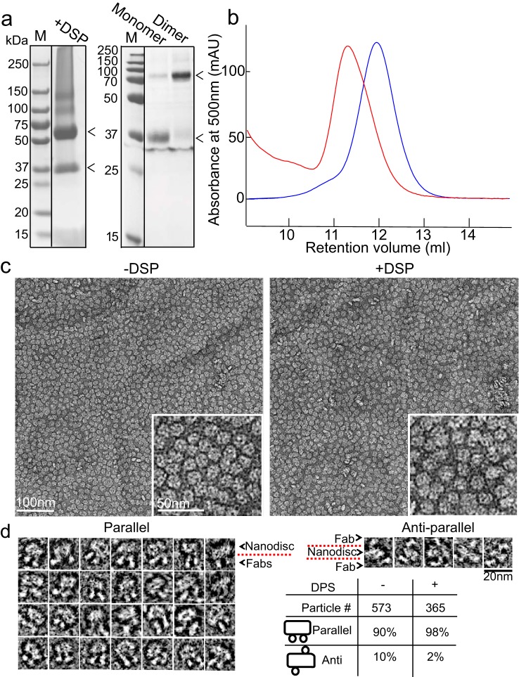
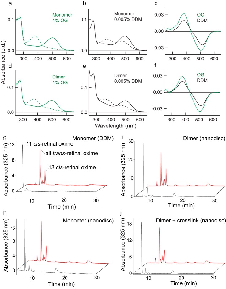
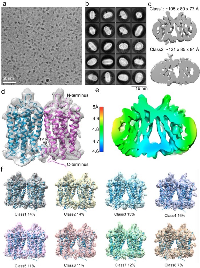
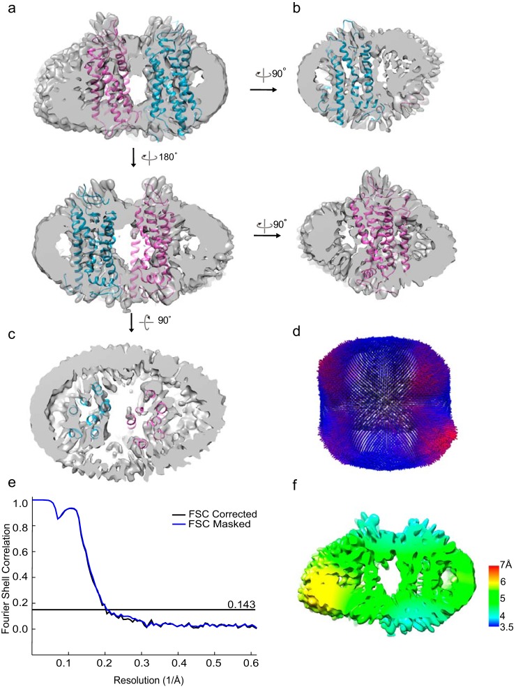
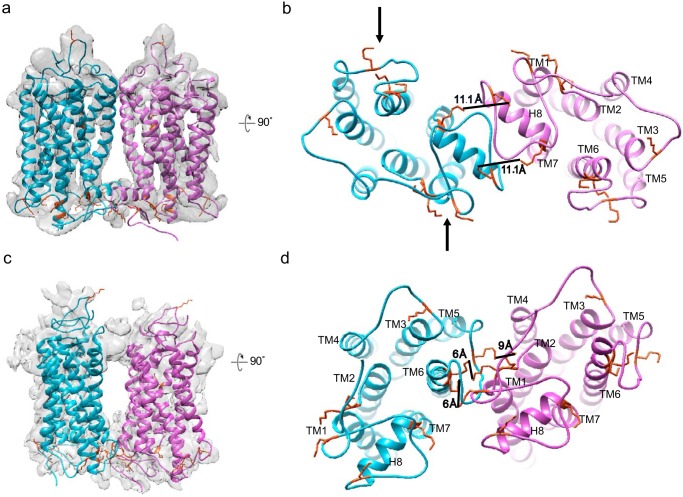
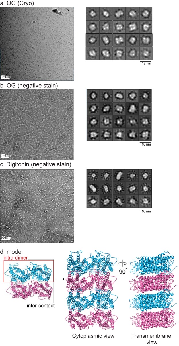
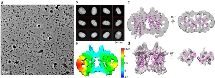
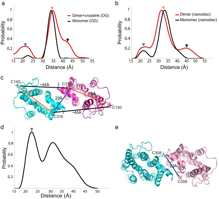
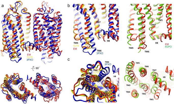
Imaging of rod photoreceptor outer-segment disc membranes by atomic force microscopy and cryo-electron tomography has revealed that the visual pigment rhodopsin, a prototypical class A G protein-coupled receptor (GPCR), can organize as rows of dimers. GPCR dimerization and oligomerization offer possibilities for allosteric regulation of GPCR activity, but the detailed structures and mechanism remain elusive. In this investigation, we made use of the high rhodopsin density in the native disc membranes and of a bifunctional cross-linker that preserves the native rhodopsin arrangement by covalently tethering rhodopsins via Lys residue side chains. We purified cross-linked rhodopsin dimers and reconstituted them into nanodiscs for cryo-EM analysis. We present cryo-EM structures of the cross-linked rhodopsin dimer as well as a rhodopsin dimer reconstituted into nanodiscs from purified monomers. We demonstrate the presence of a preferential 2-fold symmetrical dimerization interface mediated by transmembrane helix 1 and the cytoplasmic helix 8 of rhodopsin. We confirmed this dimer interface by double electron-electron resonance measurements of spin-labeled rhodopsin. We propose that this interface and the arrangement of two protomers is a prerequisite for the formation of the observed rows of dimers. We anticipate that the approach outlined here could be extended to other GPCRs or membrane receptors to better understand specific receptor dimerization mechanisms.

Keywords: G protein-coupled receptor (GPCR); cell signaling; cryo-electron microscopy (cryo-EM); double electron-electron resonance (DEER); helix 8 (H8); receptor; retina; retinoid-binding protein; rhodopsin; rhodopsin dimerization; rod outer segment; transducin; transmembrane helix 1 (TM1).


Conflict of interest statement

The authors declare that they have no conflicts of interest with the contents of this article

Figures

Figure 1.
Figure 1.
Formation of rhodopsin dimers in nanodiscs and characterization by 1D4 Fab. a, Coomassie-stained gels show that by cross-linking, primarily dimers were obtained after solubilization, before (left) or after (right) gel filtration (Fig. S1c). The arrowheads point to the positions of rhodopsin dimer and monomer on the gel. b, gel filtration of rhodopsin dimer-containing nanodiscs (red) and monomer-containing nanodiscs (blue) showing distinct peaks. c, negatively stained micrographs show that nanodiscs with in vitro reconstituted or in disc membranes cross-linked (with DSP) rhodopsin dimer are uniform in size. d, 1D4 Fab quantification of dimer orientation shows that both in disc membranes cross-linked dimers and the in vitro reconstituted dimers form parallel dimers.
Figure 2.
Figure 2.
Functional activity of rhodopsin dimer by Meta-II transition and retinoid analyses. Shown are UV-visible spectra of DSP-cross-linked rhodopsin monomer (a and b) and dimer (d and e) fractionated by gel filtration and diluted with 1.0% OG (a and d) or 0.005% DDM (b and e). Dark- and light-activated rhodopsin spectra are indicated as solid lines and dashed lines, respectively. Difference spectra (after activation − before activation) for monomer (c) and dimer (f) are shown to expand the light-dependent formation of active Meta-II (380 nm) from dark rhodopsin (500 nm). g–j, retinoid analysis shows normal 11-cis-retinal to all-trans-retinal transition upon light activation; some 13-cis-retinal is observed as a by-product in all cases. The retinals in monomer in DDM (g) or in nanodiscs (h) and dimer in nanodiscs without cross-link (i) or with cross-link (j) were converted to corresponding retinal oximes and analyzed by HPLC monitoring the UV absorbance at 325 nm. Compared with the dark state (black line), light activation (red line) shows a shift from 11-cis-retinal to all-trans-retinal.
Figure 3.
Figure 3.
Cryo-EM analysis of the rhodopsin dimer cross-linked in native disc membranes and then reconstituted into nanodiscs. a, representative cryo-EM micrograph imaged at 300 kV with the VPP in combination with a K2 detector at a pixel size of 0.8 Å and a total dose of 50 e/Å2. b, Relion 2D classification; classes are aligned to the protein density and are ranked by population. c, 762,000 particles were selected for 3D classifications; the two main refined classes have different dimensions. d, upon the removal of nanodisc EM density, docking of rhodopsin (PDB entry 1U19) into the rhodopsin EM density shows that the intradimeric interface is mediated by TM1 and H8 of each rhodopsin. e, local resolution estimation. f, class 1 particles were further classified by a second round of 3D classification (C2 symmetry) for eight classes; all analyses showed TM1 and H8 at the intradimeric interface, and no additional dimer configuration was identified. Segmented views with the docking of rhodopsin (PDB entry 1U19) are shown.
Figure 4.
Figure 4.
An interdimeric contact is observed in the cross-linked rhodopsin dimer cryo-EM data set. a–c, from 3D classification (C1 symmetry) class 2, a refined density map (C1 symmetry) with 213,000 particles was obtained. Cross-sectional views of the rhodopsin dimer with the docking of rhodopsin (PDB entry 1U19) are shown. d, angular distribution of the refined structure shows no strong bias in particle orientation for the reconstruction. e, Relion post-processing with the FSC = 0.143 criterion. f, local resolution estimation.
Figure 5.
Figure 5.
Modeling of Lys ϵ-amino groups as cross-link sites in the intradimeric interface and the interdimeric contact. A total of nine exposed Lys residues on the cytoplasmic side of rhodopsin are available for chemical cross-linking by DSP. Black arrows point to Lys residues that can be linked to DSP and thus block Gt interaction. Shown are side (a) and cytoplasmic (b) views of the intradimeric interface with rhodopsin crystal structure (PDB entry 1U19) docked into the EM densities. Lys-311 and Lys-325 on both rhodopsin molecules allow two cross-links to form. The cross-sectional area taken up by intradimer is 84 × 47 Å = 3948 Å2. Shown are side (c) and cytoplasmic (d) views of the interdimeric contact; Lys-245/248/231 on one rhodopsin can cross-link with Lys-66/67 on the adjacent rhodopsin based on the 12-Å length of the DSP linker. The cross-sectional area of the interdimer is 87 × 64 Å = 5568 Å2. The MSP1E3D1 nanodisc is relatively small for the observed large cross-sectional area and could result in some distortion of the rhodopsin arrangement.
Figure 6.
Figure 6.
Cross-linked rhodopsin tetramer structure and model of rhodopsin oligomers. a, rhodopsin cross-linked in disc membranes was solubilized in 2% OG, and tetramers were isolated by gel filtration, vitrified, and imaged by cryo-EM with a Tecnai F20. Particles (∼65,000) were autopicked and analyzed for 2D classification by Relion 2. Most of the tetramer classes have a square shape rather than linear, T, or L shape. A representative micrograph is shown to the left. Cross-linked tetramers solubilized in 2% OG (b) or in 0.1% digitonin (c) were negatively stained and imaged with a Tecnai F20, and 35,000 particles from each condition were autopicked and analyzed by Relion 2. Most of the tetramer classes have a square shape. A representative micrograph is shown to the left. Some 2D classes for hexamer are also seen with 0.1% digitonin solubilization. Circle, represents mask diameter at 180 Å. d, model of rhodopsin oligomer. A pair of rhodopsin molecules with the same color forms the intradimeric interface, whereas two adjacent rhodopsin molecules with different colors form the interdimeric contact. The organization of the tetramer and oligomer (cytoplasmic and transmembrane views) is displayed.
Figure 7.
Figure 7.
Cryo-EM analysis of the in vitro reconstituted rhodopsin dimer in nanodiscs. a, representative cryo-EM micrograph imaged at 300 kV using the VPP in combination with a K2 detector at a pixel size of 1.35 Å and a total dose of 50 e/Å2. b, Relion 2D classification; classes are ranked by population; classes with the transmembrane view are boxed in red. c, 3D refinement (C2 symmetry) of 96,000 particles aligned to protein densities shows that the intradimeric interface is mediated by TM1 and H8; two rhodopsin molecules (PDB entry 1U19) are docked, with cross-sectional views shown from the side (left) and bottom (right). d, docking of rhodopsin (PDB entry 1U19) into the EM density after nanodisc removal in Chimera; cross-sectional view shown to the right. e, local resolution estimate (C2 symmetry).
Figure 8.
Figure 8.
DEER distance distributions of spin-labeled rhodopsin dimer. a, rhodopsin monomer (black) with Cys-140 and Cys-316 spin-labeled in 3% OG showed a single peak centered at ∼35 Å. In disc membranes, cross-linked rhodopsin dimer (red) in 3% OG showed additional peaks at ∼23 and ∼45 Å. b, rhodopsin monomer reconstituted into nanodiscs (black) with Cys-140 and Cys-316 spin-labeled showed a peak centered ∼35 Å. In vitro reconstituted rhodopsin dimer in nanodiscs (red) showed additional peaks at ∼23 and ∼45 Å. c, schematics of the rhodopsin dimer configuration predicted by the DEER measurement, with the distances between Cys-140 and Cys-316 color-labeled. The nitroxide label is modeled into the Cys-140 and Cys-316 sites of rhodopsin. d, rhodopsin mutant spin-labeled at site 308 and in vitro reconstituted as dimer in nanodisc showed additional intradimeric DEER distance at ∼23 Å (blue arrow); the width of the peak suggests flexibility of the intradimeric interface. e, schematic of the dimer configuration predicted by the DEER measurement, with the distance from Cys-308 labeled. The nitroxide label is modeled into the Cys-308 site of the rhodopsin mutant.
Figure 9.
Figure 9.
Comparison of rhodopsin cryo-EM structure with known GPCR class A crystal structures. Overlay of EM rhodopsin dimer structure (red) with crystal structures of dark state rhodopsin monomer (yellow, PDB entry 1U19), active opsin dimer (blue, PDB entry 3PXO), and inactive β1-AR dimer (green, PDB entry 4GPO). a, top, side view; bottom, cytoplasmic view. b and c, enlarged side view and cytoplasmic view of TM5/TM6 regions that change upon rhodopsin activation. Left, EM rhodopsin dimer superimposed with inactive rhodopsin (PDB entry 1U19) and activated opsin (PDB entry 3PXO). Right, EM rhodopsin dimer superimposed with β1-AR (PDB entry 4GPO).
Figure 10.
Figure 10.
Overlay of rhodopsin–Gi and rhodopsin–arrestin complex structures with rhodopsin docked into intradimeric EM densities (Fig. 3d). Shown are a side view (a) and cytoplasmic view (b) of activated rhodopsin (blue); and two Gαβγ complex: one in yellow and one in orange. Two Gi complexes are separated by a dotted line. Shown are a side view (c) and cytoplasmic view (d) of activated rhodopsin (blue); and two arrestin: one in magenta and one in cyan, separated by a dotted line. Shown are a side view (e) and cytoplasmic view (f) of activated rhodopsin (blue), a G complex in orange, and the arrestin in cyan. A dotted line separates arrestin from the Gi complex.

References

    1. Salon J. A., Lodowski D. T., and Palczewski K. (2011) The significance of G protein-coupled receptor crystallography for drug discovery. Pharmacol. Rev. 63, 901–937 10.1124/pr.110.003350 - DOI - PMC - PubMed
    1. Reiter E., Ahn S., Shukla A. K., and Lefkowitz R. J. (2012) Molecular mechanism of β-arrestin-biased agonism at seven-transmembrane receptors. Annu. Rev. Pharmacol. Toxicol. 52, 179–197 10.1146/annurev.pharmtox.010909.105800 - DOI - PMC - PubMed
    1. Rohrer D. K., and Kobilka B. K. (1998) G protein-coupled receptors: functional and mechanistic insights through altered gene expression. Physiol. Rev. 78, 35–52 10.1152/physrev.1998.78.1.35 - DOI - PubMed
    1. Palczewski K., Kumasaka T., Hori T., Behnke C. A., Motoshima H., Fox B. A., Le Trong I., Teller D. C., Okada T., Stenkamp R. E., Yamamoto M., and Miyano M. (2000) Crystal structure of rhodopsin: a G protein-coupled receptor. Science 289, 739–745 10.1126/science.289.5480.739 - DOI - PubMed
    1. Palczewski K. (2006) G protein-coupled receptor rhodopsin. Annu. Rev. Biochem. 75, 743–767 10.1146/annurev.biochem.75.103004.142743 - DOI - PMC - PubMed

Publication types