. 2020 Jan 17;295(3):743-756.
doi: 10.1074/jbc.RA119.010188. Epub 2019 Dec 10.

The structure of the Thermococcus gammatolerans McrB N-terminal domain reveals a new mode of substrate recognition and specificity among McrB homologs

Affiliations

The structure of the Thermococcus gammatolerans McrB N-terminal domain reveals a new mode of substrate recognition and specificity among McrB homologs

Christopher J Hosford et al. J Biol Chem. .

Abstract

McrBC is a two-component, modification-dependent restriction system that cleaves foreign DNA-containing methylated cytosines. Previous crystallographic studies have shown that Escherichia coli McrB uses a base-flipping mechanism to recognize these modified substrates with high affinity. The side chains stabilizing both the flipped base and the distorted duplex are poorly conserved among McrB homologs, suggesting that other mechanisms may exist for binding modified DNA. Here we present the structures of the Thermococcus gammatolerans McrB DNA-binding domain (TgΔ185) both alone and in complex with a methylated DNA substrate at 1.68 and 2.27 Å resolution, respectively. The structures reveal that TgΔ185 consists of a YT521-B homology (YTH) domain, which is commonly found in eukaryotic proteins that bind methylated RNA and is structurally unrelated to the E. coli McrB DNA-binding domain. Structural superposition and co-crystallization further show that TgΔ185 shares a conserved aromatic cage with other YTH domains, which forms the binding pocket for a flipped-out base. Mutational analysis of this aromatic cage supports its role in conferring specificity for the methylated adenines, whereas an extended basic surface present in TgΔ185 facilitates its preferential binding to duplex DNA rather than RNA. Together, these findings establish a new binding mode and specificity among McrB homologs and expand the biological roles of YTH domains.

Keywords: 6-methyladenosine; DNA binding; DNA binding protein; McrB; RNA-protein interaction; X-ray crystallography; YTH domain; protein structure; protein-nucleic acid interaction; restriction system; structural biology.


Conflict of interest statement

The authors declare that they have no conflicts of interest with the contents of this article

Figures

Figure 1.
Figure 1.
N-terminal domains of McrB homologs are not conserved. The diagram illustrates phylogenetic analysis of representative McrB homologs. Conserved C-terminal, GTP-specific AAA+ domains are colored light blue. Divergent N-terminal domains are colored differently according to the predicted fold. The protein folds of homologs highlighted in red have been experimentally validated by X-ray crystallography. Department of Energy Integrated Microbial Genomes codes (62) and any applicable PDB codes are as follows: Yersinia pestis sv. Orientalis CO-92 McrB, 637199492; Acinetobacter baumannii D1279779 McrB, 2563734192; Bacillus cereus 03BB102 McrB, 643761466; Thermococcus gammatolerans EJ3 McrB, 644807740; Staphylococcus aureus MRSA252 McrB, 637153557; Lysinibacillus fusiformis SW-B9 McrB, 2598933124; Firmicutes bacterium JGI 0000119-P10 McrB, 2519130374; Rhizobium sp. CF097 McrB, 2585392831; E. coli K-12 MG1655 McrB, 646316336, PDB code 3SSC; Staphylothermus marinus F1, DSM 3639 McrB, 640109242, PDB code 6N0S; Lactococcus lactis lactis 1AA59 LlaI.1, 263206860; L. lactis lactis 1AA59 LlaI.2, 2632068606; L. lactis LlaJI.R1, 642916737; and H. pylori LlaJI.R1, 637022177, PDB code 6C5D.
Figure 2.
Figure 2.
TgΔ185 binds m5C dsDNA. A and B, domain architectures of EcMcrB (A) and TgMcrB (B) N-terminal domains. Ec DNA-binding domain (EcΔ155) is colored orange and Tg N-terminal domain (TgΔ185) is colored yellow. The conserved C-terminal AAA+ domain is colored light blue. Truncated constructs used for crystallization and SEC experiments are indicated by the dashed boxes. C, size shift of EcΔ155 (upper panel) and EcΔ155 + m5C dsDNA (lower panel) are visualized for change in retention volume off of SEC on SDS-PAGE gels silver-stained for DNA and Coomassie-stained for protein. D, size shift of TgΔ185 (upper panel) and TgΔ155 + m5C dsDNA (lower panel) are visualized for change in retention volume off of SEC on SDS-PAGE gels silver-stained for DNA and Coomassie-stained for protein. EcΔ155 and TgΔ185 are both capable of binding the same m5C DNA substrates as indicated by the respective protein bands size shift to an earlier retention volume. E, filter-binding analysis of TgMcrB and EcMcrB binding to 5-methylctosine modified (m5C) and nonmethylated (nm) dsDNA substrates. See Table S1 for substrate sequences. The data points represent the averages of at least three independent experiments (means ± S.D.). Binding constants were determined by nonlinear curve fitting using Kaleidagraph (Synergy Software) and defined as the concentration of the protein at which 50% of the labeled DNA substrate is retained. Calculated Kd values are listed in Table 1.
Figure 3.
Figure 3.
TgΔ185 adopts a YTH fold that is distinct from the EcΔ155 fold. A and B, structure (A) and topology (B) of TgΔ185 (yellow). C and D, structure (C) and topology (D) of EcΔ155 (orange). E, topology diagram of HsYTHDF2 YTH domain (light blue). F, structural superposition of TgΔ185 (yellow) and the HsYTHDF2 YTH domain (light blue).
Figure 4.
Figure 4.
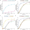
TgMcrB preferentially binds DNA containing m6A modifications. All data points represent average of three independent experiments (means ± S.D.). Binding constants were determined by nonlinear curve fitting using Kaleidagraph (Synergy Software) and defined as the concentration of the protein at which 50% of the labeled DNA substrate is retained. Substrate sequences and calculated Kd values are listed in Table S1 and Table 1, respectively. m5C and m6A denote 5-methylcytosine and 6-methyladenine modifications, respectively. nmC and nmA denote nonmethylated versions of the same substrates. A, filter-binding analysis of TgMcrB and HsYTHDC1 YTH domain interactions with RNA substrates. B, filter-binding analysis of TgMcrB interactions with dsDNA substrates. Binding curves from Fig. 2E are included for comparison. C, filter-binding analysis of TgMcrB interaction with different single stranded DNA (ssDNA) substrates. D, filter-binding analysis of TgMcrB with different mismatched dsDNA substrates.
Figure 5.
Figure 5.
DNA-bound structure of TgΔ185. A, cartoon representation of TgΔ185 bound to m5C-containing, mismatched dsDNA substrate with mismatches (meDNA; Table S1) shown in two orientations. TgΔ185 is colored yellow, and bound DNA is colored wheat. B, schematic of the meDNA substrate used for crystallization with TgΔ185. Mismatched bases are colored red and indicated by arrows. C, crystal packing of TgΔ185 with meDNA. One asymmetric unit is colored yellow with the bound DNA illustrated as sticks and colored wheat. The electron density map associated with the DNA is colored light gray and illustrated as mesh. D, zoomed-in view of the electron density surrounding the flipped-out adenine base. E, zoomed-in view of the electron density surrounding base pairs within the bound DNA duplex. F, structural comparison of meDNA with B-form DNA (PDB code 1bna) illustrates deformation in the bound DNA.
Figure 6.
Figure 6.
TgΔ185 utilizes a structurally conserved aromatic cage to bind DNA. A, zoomed-in view of the TgΔ185 aromatic cage residues (yellow) and modeled adenine base from co-crystallized DNA substrate (wheat). B, zoomed in view of the HsYTHDC1 aromatic cage residues (green) with bound m6A base from co-crystallized RNA substrate (wheat; PDB code 4r3i). C, filter-binding analysis of TgMcrB WT and aromatic cage mutants with m6A dsDNA (see Table S1 for sequence). The data points represent averages of three independent experiments (means ± S.D.). Binding constants were determined by nonlinear curve fitting using Kaleidagraph (Synergy Software) and defined as the concentration of the protein at which 50% of the labeled DNA substrate is retained. Calculated Kd values are listed in Table 1. D and E, electrostatic surfaces of HsYTHDC1 with bound m6A-modified ssRNA substrate (PDB code 4r3i; D and TgΔ185 with bound mismatched dsDNA substrate (E). A yellow box is drawn around the position of the aromatic cage in both structures and indicated by arrows. The scale bar indicates electrostatic surface coloring from −3 KbT/ec to +3 KbT/ec.
Figure 7.
Figure 7.
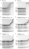
EMSA analysis of predicted TgMcrBΔ185 DNA-binding mutants. Binding was carried out at 25 °C for 30 min in a 16-μl reaction mixture containing 5 ng/μl of digested (BamHI/NdeI) m6A methylated (dam+) and nonmethylated (dam) λ-phage DNA and increasing concentrations (0–10 μm) of each full-length TgMcrB construct. The gels were stained with SYBR® Green in 1× TAE overnight at 25 °C. Calculated sizes (bp) of the digested DNA products are noted on the left of each gel.

References

    1. Labrie S. J., Samson J. E., and Moineau S. (2010) Bacteriophage resistance mechanisms. Nat. Rev. Microbiol. 8, 317–327 10.1038/nrmicro2315 - DOI - PubMed
    1. Loenen W. A., and Raleigh E. A. (2014) The other face of restriction: modification-dependent enzymes. Nucleic Acids Res. 42, 56–69 10.1093/nar/gkt747 - DOI - PMC - PubMed
    1. Bair C. L., and Black L. W. (2007) A type IV modification dependent restriction nuclease that targets glucosylated hydroxymethyl cytosine modified DNAs. J. Mol. Biol. 366, 768–778 10.1016/j.jmb.2006.11.051 - DOI - PMC - PubMed
    1. Borgaro J. G., and Zhu Z. (2013) Characterization of the 5-hydroxymethylcytosine-specific DNA restriction endonucleases. Nucleic Acids Res. 41, 4198–4206 10.1093/nar/gkt102 - DOI - PMC - PubMed
    1. Ishikawa K., Fukuda E., and Kobayashi I. (2010) Conflicts targeting epigenetic systems and their resolution by cell death: novel concepts for methyl-specific and other restriction systems. DNA Res. 17, 325–342 10.1093/dnares/dsq027 - DOI - PMC - PubMed

Publication types

MeSH terms