. 2019 Sep 12;2(5):e201900458.
doi: 10.26508/lsa.201900458. Print 2019 Oct.

Structural and functional characterization of the mitochondrial complex IV assembly factor Coa6

Affiliations

Structural and functional characterization of the mitochondrial complex IV assembly factor Coa6

Shadi Maghool et al. Life Sci Alliance. .

Abstract

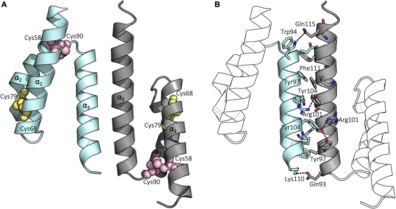
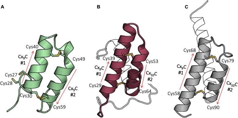
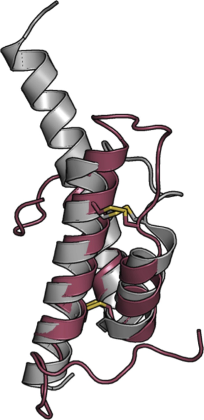
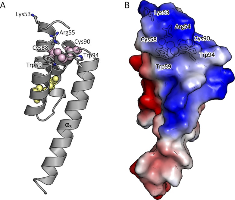
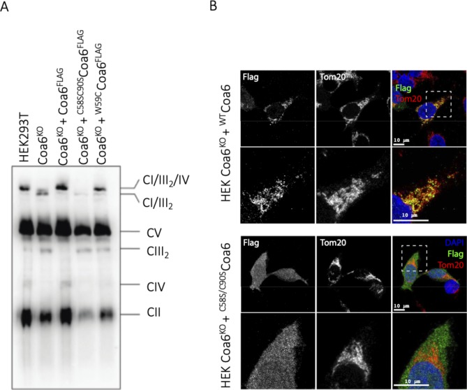
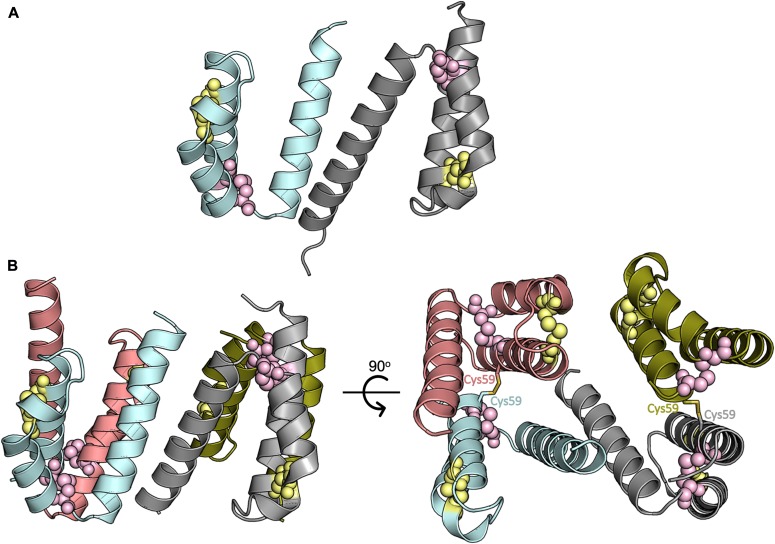
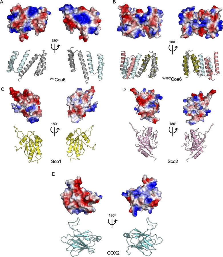
Assembly factors play key roles in the biogenesis of many multi-subunit protein complexes regulating their stability, activity, and the incorporation of essential cofactors. The human assembly factor Coa6 participates in the biogenesis of the CuA site in complex IV (cytochrome c oxidase, COX). Patients with mutations in Coa6 suffer from mitochondrial disease due to complex IV deficiency. Here, we present the crystal structures of human Coa6 and the pathogenic W59CCoa6-mutant protein. These structures show that Coa6 has a 3-helical bundle structure, with the first 2 helices tethered by disulfide bonds, one of which likely provides the copper-binding site. Disulfide-mediated oligomerization of the W59CCoa6 protein provides a structural explanation for the loss-of-function mutation.


Conflict of interest statement

The authors declare that they have no conflict of interest.

Figures

Figure 1.
Figure 1.. Structure of the WTCoa6.
(A) Cartoon representation of the overall structure of WTCoa6. Secondary structures are represented as cartoons with monomers colored in cyan and gray. Residues Cys58 and Cys90 are shown as pink spheres, whereas Cys68 and Cys79 residues are shown as yellow spheres. (B) Residues located at the dimer interface are shown as sticks and hydrogen bonds between residues (labeled) shown as dashed lines.
Figure 2.
Figure 2.. WTCoa6 has a twin CX9C protein fold.
(A) Cox17 (PDB 2RNB): helices are shown in green, and cysteines in the CX9C motifs that form disulfide bonds are shown as yellow sticks. (B) Cox6B (PDB 2EIJ): helices are shown in raspberry, and cysteines in the CX9C (or CX10C) motifs that form disulfide bonds are shown as yellow sticks. (C) WTCoa6 (this work): helices are shown in gray, and cysteines in the CX9C (or CX10C) motifs that form disulfide bonds are shown as yellow sticks. In all panels, the positions of the corresponding motifs are shown as red arrows. In panel (B), part of the N terminus and helix α3 in panels (B) and (C) are not colored for clarity.
Figure S1.
Figure S1.. Comparison of the WTCoa6 (gray) and COX6B (raspberry) structures.
WTCoa6 shares significant structural similarity with the Cox6B subunit of COX (PDB 2ZXW, chains H and U) (31).
Figure S2.
Figure S2.. Analytical SEC analysis of WTCoa6 and mutant Coa6 proteins.
All the mutated proteins eluted at a volume similar to the WTCoa6 dimeric form from an analytical size-exclusion column. Elution profiles for the WTCoa6, R101ACoa6, Y97ACoa6, Y104ACoa6, and Y97A/Y104ACoa6 proteins are shown as blue, green, black, red, and gray lines, respectively.
Figure S3.
Figure S3.. Determination of copper content of copper-loaded WTCoa6.
A standard curve (top) was used to determine the copper content of the WTCoa6 (bottom).
Figure S4.
Figure S4.. Redox potential measurements for WTCoa6.
SDS–PAGE analysis of WTCoa6 (200 μM) incubated for 2 h with increasing concentrations of reduced DTT (0–800 mM) followed by AMS labeling. The reduced fractions were quantified using the ImageJ software (59). The fraction of thiolate as a function of [DTTRed]/[DTTOx] is plotted to calculate the equivalent intrinsic redox potential (−349 ± 1 mV for WTCoa6 at pH 7.0).
Figure S5.
Figure S5.. WTCoa6 mass determined via MALDI-TOF following reduction and IAA labeling.
(A) The dominant species in the oxidized WTCoa6 sample included no free thiol groups (2S-S). (B) The dominant species in the reduced WTCoa6 sample included two free thiol groups (2SH [not 4SH]).
Figure 3.
Figure 3.. Determination of KD for Cu(I) binding for the WTCoa6, C58S/C90SCoa6, and C68S/C79SCoa6 proteins.
The data were analyzed via a plot of the concentration of the [CuI(BCS)2]3− complex (calculated from the absorption values at 483 nm versus the protein:Cu ratio) and the data fit using the equation previously described (11, 35).
Figure S6.
Figure S6.. Secondary structure analyses of the WTCoa6, C58S/C90SCoa6, and C68S/C79SCoa6-mutant proteins.
CD spectra for WTCoa6 (solid circles), C58S/C90SCoa6 (circles), and C68S/C79SCoa6 (diamonds) at 0.16 mg·ml−1 and at 20°C. CD spectra show minima at 222 and 208 nm characteristic of α-helical structures.
Figure S7.
Figure S7.. Positively charged residues in the vicinity of the Cu(II)-binding site in WTCoa6.
(A) Cartoon representation of the chain B of WTCoa6. The structure of WTCoa6 shows that the Cys58–Cys90 site is proximate to positively charged residues Lys53 and Arg55, which are shown as sticks (labeled) and bordered by the aromatic side chains of residues Trp59 and Trp94 (sticks, labeled), which partially shield the site from solvent. (B) Surface representation of chain B of WTCoa6. The protein surfaces are colored according to their electrostatic potentials (red, negatively charged; blue, positively charged; and white, uncharged). Cysteine residues are shown as spheres for clarity.
Figure S8.
Figure S8.. Loss of Coa6 results in complex IV deficiency.
(A) Cells were solubilized in digitonin and subjected to BN-PAGE and immunoblot analysis using a total OXPHOS antibody cocktail. SC, supercomplexes consisting of CI, CIII, and CIV. (B) Unlike WTCoa6, the C58S/C90SCoa6-mutant localizes in the cytosol. The FLAG-tagged WTCoa6 and FLAG-tagged C58S/C90SCoa6 double mutant were expressed in COA6KO cells. Immunofluorescence was performed using Flag and Tom20 antibodies. The resulting images were merged using ImageJ software. The scale bar represents 10 μm.
Figure 4.
Figure 4.. Structure of the W59CCoa6-mutant protein.
(A) Cartoon representation of the overall structure of WTCoa6. Secondary structures are represented as cartoons with monomers colored in cyan and gray. (B) Cartoon representation of the overall structure of W59CCoa6. Secondary structures are represented as cartoons with monomers colored in cyan, gray, salmon and gold. Residues Cys58 and Cys90 are shown as pink spheres and residues Cys68 and Cys79 as yellow spheres. Each monomer (cyan and gray) is linked to another monomer (salmon and gold, respectively) by an intermolecular disulfide bond (shown as yellow sticks) through the introduced Cys59 residue.
Figure S9.
Figure S9.. Surface electrostatic patterns of WTCoa6, W59CCoa6, Sco1, Sco2, and soluble domain of COX2 proteins.
(A) WTCoa6. (B) W59CCoa6. (C) Sco1 (PDB 2GVP) (70). (D) Sco2 (PDB 2RLI) (9). (E) Soluble domain of human COX2 (PDB 5Z62) (71). Top panels: the molecular surface of proteins is colored according to the electrostatic potentials (red, negatively charged; blue, positively charged; and white, uncharged). Bottom panels: secondary structures are represented as cartoons as in the top panels. WTCoa6 monomers colored in cyan and gray; W59CCoa6 monomers colored in cyan, gray, orange, and green; Sco1 colored in yellow; Sco2 colored in pink and soluble domain of COX2 colored in cyan.

References

    1. Ferguson-Miller S, Babcock GT (1996) Heme/copper terminal oxidases. Chem Rev 96: 2889–2908. 10.1021/cr950051s - DOI - PubMed
    1. Soto IC, Fontanesi F, Liu J, Barrientos A (2012) Biogenesis and assembly of eukaryotic cytochrome c oxidase catalytic core. Biochim Biophys Acta 1817: 883–897. 10.1016/j.bbabio.2011.09.005 - DOI - PMC - PubMed
    1. Fernandez-Vizarra E, Tiranti V, Zeviani M (2009) Assembly of the oxidative phosphorylation system in humans: What we have learned by studying its defects. Biochim Biophys Acta 1793: 200–211. 10.1016/j.bbamcr.2008.05.028 - DOI - PubMed
    1. Timon-Gomez A, Nyvltova E, Abriata LA, Vila AJ, Hosler J, Barrientos A (2018) Mitochondrial cytochrome c oxidase biogenesis: Recent developments. Semin Cell Dev Biol 76: 163–178. 10.1016/j.semcdb.2017.08.055 - DOI - PMC - PubMed
    1. Tsukihara T, Aoyama H, Yamashita E, Tomizaki T, Yamaguchi H, Shinzawa-Itoh K, Nakashima R, Yaono R, Yoshikawa S (1996) The whole structure of the 13-subunit oxidized cytochrome c oxidase at 2.8 A. Science 272: 1136–1144. 10.1126/science.272.5265.1136 - DOI - PubMed

Publication types

LinkOut - more resources