. 2019 Oct 11;294(41):14845-14859.
doi: 10.1074/jbc.RA119.010572. Epub 2019 Sep 5.

CD23 is a glycan-binding receptor in some mammalian species

Affiliations

CD23 is a glycan-binding receptor in some mammalian species

Sabine A F Jégouzo et al. J Biol Chem. .

Abstract

CD23, the low-affinity IgE receptor found on B lymphocytes and other cells, contains a C-terminal lectin-like domain that resembles C-type carbohydrate-recognition domains (CRDs) found in many glycan-binding receptors. In most mammalian species, the CD23 residues required to form a sugar-binding site are present, although binding of CD23 to IgE does not involve sugars. Solid-phase binding competition assays, glycoprotein blotting experiments, and glycan array analysis employing the lectin-like domains of cow and mouse CD23 demonstrate that they bind to mannose, GlcNAc, glucose, and fucose and to glycoproteins that bear these sugars in nonreducing terminal positions. Crystal structures of the cow CRD in the presence of α-methyl mannoside and GlcNAcβ1-2Man reveal that a range of oligosaccharide ligands can be accommodated in an open binding site in which most interactions are with a single terminal sugar residue. Although mouse CD23 shows a pattern of monosaccharide and glycoprotein binding similar to cow CD23, the binding is weaker. In contrast, no sugar binding was observed in similar experiments with human CD23. The absence of sugar-binding activity correlates with accumulation of mutations in the gene for CD23 in the primate lineage leading to humans, resulting in loss of key sugar-binding residues. These results are consistent with a role for CD23 in many species as a receptor for potentially pathogenic microorganisms as well as IgE. However, the ability of CD23 to bind several different ligands varies between species, suggesting that it has distinct functions in different organisms.

Keywords: CLEC4J; FCER2 gene; Fc receptor; carbohydrate function; carbohydrate-binding protein; carbohydrate-recognition domain; crystal structure; glycan-binding receptors; glycobiology; lectin.


Conflict of interest statement

The authors declare that they have no conflicts of interest with the contents of this article

Figures

Figure 1.
Figure 1.
Expression of cow CD23. A, domain organization of CD23. B, sequence alignment of CD23 from human, mouse, and cow. The transmembrane region is highlighted in violet, and the a and d positions of the heptad repeats in the neck are highlighted in orange. Conserved cysteine residues, linked by disulfide bonds, are shaded yellow. Positions of residues that commonly interact with Ca2+ are indicated in green for the conserved sugar-binding site and in pink for an accessory site found in some C-type CRDs. Residue numbers correspond to the cow protein based on sequence XP_002688905.2 from the National Center for Biotechnology Information. C and D, SDS-PAGE of fractions eluted from a mannose-Sepharose column with EDTA for the CRD fragment alone and with an appended biotin tag. Gels were stained with Coomassie Blue.
Figure 2.
Figure 2.
Characterization of cow CD23 binding to sugars in a solid-phase binding assay. A, binding competition assays in which immobilized CRD was probed with horseradish peroxidase, as reporter ligand, at concentrations well below saturation binding. Under these conditions, the relative KI values for competing monosaccharide and oligosaccharide ligands closely approximate KD values. Solid bars, KI values for various sugars relative to the KI for mannose. Error bars, S.D. for three or more assays for each sugar. Absolute KI values are indicated at the top of each bar. B, comparison of binding of α-methyl mannoside and Man9GlcNAc2 oligosaccharide. An example of a competition assay is shown. The average ratio of the affinities for five assays was 240 ± 50. C, pH dependence of binding was determined using the same solid-phase assay format.
Figure 3.
Figure 3.
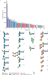
Glycan array analysis of cow CD23. A, a glycan array comprising oligosaccharides from the Consortium for Functional Glycomics was probed with a tetravalent complex of biotin-tagged CRD with fluorescently labeled streptavidin. Results are color-coded based on the presence of reducing-end GlcNAcβ1–2Man epitopes (blue bars), GlcNAcβ1–2Man with galactose linked 1–3 to the GlcNAc residue (cyan bars), oligomannose structures (green bars), or Fucα1–2Gal disaccharide (red bars). Complete results for all oligosaccharides on the array are given in Table S1. B, structures of oligosaccharide ligands that show the strongest signals on the glycan array. Error bars, S.D.
Figure 4.
Figure 4.
Probing of glycoproteins with cow CD23. Glycoproteins were separated by SDS-PAGE. In each panel, the Coomassie Blue–stained gel is shown on the left, and a blot probed with CRD-avidin-alkaline phosphatase complex is shown on the right. A, serum glycoproteins. B, natural glycoproteins and versions that have been modified to expose novel reducing-end sugars.
Figure 5.
Figure 5.
Structure of the CRD from cow CD23. A, overall structure of the CRD bound to GlcNAcβ1–2Man. The protein in a cartoon representation is colored from blue (N terminus) to red (C terminus). Disulfide bonds are shown in yellow. The disaccharide ligand is shown in a stick representation, with carbon atoms in gray, oxygen atoms in red, and Ca2+ in orange. B, superposition of ligand-binding sites in the three crystal structures, with carbon atoms shown in yellow for α-methyl mannoside, gray for GlcNAcβ1–2Man, and cyan for GlcNAc2Man3. Oxygen, nitrogen, and Ca2+ are as in A. C–E, FoFc electron density maps calculated by omitting the sugar residue from the model, contoured at 3.0 σ, and shown as a green mesh for α-methyl mannoside (C), GlcNAcβ1–2Man (D), and GlcNAc2Man3 (E). F–I, close-up views of the Ca2+ and the ligand-binding sites. F, α-methyl mannoside in the primary sugar-binding site, with OH groups 3 and 4 of mannose ligated to the conserved Ca2+. For clarity, only one orientation of the sugar is shown. G, accessory Ca2+ site, with ligands from four side chains as well as the backbone carbonyl group of Glu259. H, GlcNAcβ1–2Man in the primary sugar-binding site, ligated to the conserved Ca2+. I, interactions of the GlcNAc moiety of GlcNAcβ1–2Man at the secondary binding site. The protein in cartoon representation as well as carbon atoms in stick representations are presented in gray, carbon atoms of the sugar are cyan for α-methyl mannoside and yellow for GlcNAcβ1–2Man, and other atoms are as in A.
Figure 6.
Figure 6.
Comparison of cow and human CD23. A, flexibility in loops 1 and 4 shown in superposition of the CRDs of cow and human CD23. The CRD from the complex of cow CD23 with α-methyl mannoside is shown in gray. Human CD23 without bound Ca2+ (PDB entries 2H2R and 4G96) is shown in blue and cyan, and CD23 with Ca2+ bound is shown in red (PDB entry 2H2T). B, superposition of the CRD of cow CD23, with GlcNAcβ1–2Man in red, human CD23 complexed with domains Cϵ3 and Cϵ4 of IgE in the presence of Ca2+ (PDB entry 4GKO) in blue, human CD23 complexed with domains Cϵ3 and Cϵ4 of IgE in the absence of Ca2+ (PDB entry 4EZM) in cyan, and CD23 complexed with domains Cϵ2–Cϵ4 of IgE in the absence of Ca2+ (PDB entry 5LGK) in green.
Figure 7.
Figure 7.
Cross-linking of two CRDs of cow CD23 complexed with a biantennary ligand. Overall orientation of CRDs bound to the two branches of the bi-antennary glycan is shown in the context of two receptor trimers. Individual terminal GlcNAcβ1–2Man disaccharides on two branches of the oligosaccharide interacting with sugar-binding sites in CRDs from CD23 and a symmetry-related molecule. The protein is shown in cartoon representation in gray and cyan. Ca2+ is represented in orange spheres. The biantennary ligand is shown in a stick representation, with carbon atoms in gray or cyan and oxygen atoms in red. The 2-fold axis relating the CRDs is perpendicular to the membrane.
Figure 8.
Figure 8.
Flow cytometry to demonstrate expression of cow CD23 on B cells. Peripheral blood mononuclear cells were isolated from cow blood and stained with monoclonal anti-CD21 and anti-CD23 antibodies. A, gating of mature B cells based on forward and side scattering and CD21 expression. B, comparison of binding of antibody to CD23 and isotype control to the B cell population.
Figure 9.
Figure 9.
Expression of mouse CD23. A, SDS-PAGE of fractions eluted from a 10-ml mannose-Sepharose column for the CRD fragment of mouse CD23 with appended biotin tag. Following application of renatured protein, the column was washed with 10 1-ml aliquots of buffer containing 25 mm Ca2+ and eluted with 15 1-ml aliquots of buffer containing 2.5 mm EDTA. Fractions 11–12, corresponding to material weakly bound to the column, were used for coating of assay plates and formation of streptavidin complexes. B, SDS-PAGE of fractions eluted from a 1-ml mannose-Sepharose column for the biotin-tagged complexed with Alexa Fluor 647–labeled streptavidin. The column was washed with 1 ml of buffer containing 25 mm Ca2+ and eluted with seven aliquots (0.25 ml) of buffer containing 2.5 mm EDTA. Gels were stained with Coomassie Blue.
Figure 10.
Figure 10.
Characterization of mouse CD23 binding to monosaccharides in solid-phase binding assay. Binding competition assays were performed with immobilized CRD probed with horseradish peroxidase. Error bars, S.D. for three or more assays for each sugar. Absolute KI values are indicated at the top of each bar.
Figure 11.
Figure 11.
Probing of glycoproteins with mouse CD23. Glycoproteins were separated by SDS-PAGE. In each panel, Coomassie Blue-stained gel is shown on the left, and the blot probed with the CRD-avidin-alkaline phosphatase complex is shown on the right. A, natural glycoproteins. B, glycoproteins modified to expose novel reducing-end sugars.
Figure 12.
Figure 12.
Evolution of Ca2+- and sugar-binding sites in CD23. A, monophyletic arrangement of groups of primates in which one or more of the residues needed to form functional Ca2+- and sugar-binding sites are mutated. For each genus, a green check mark indicates conservation of the sites compared with cow CD23, whereas a red × indicates that there are changes in one or more residues in these sites. B, sequence alignment showing details of amino acid changes in representative species within each genus. Sequences for all of the available mammalian CD23 orthologues are given in Fig. S1. Residues that ligate the conserved Ca2+ and sugar are indicated in green, and residues forming an accessory Ca2+ binding site are indicated in pink.
Figure 13.
Figure 13.
Multiple binding interfaces of CD23. Two views of the cow CD23 structure highlighting residues that interact with various ligands. A, view of the CRD with the disaccharide binding site exposed. B, view of the opposite face of the CRD. Red, residues that bind to sugars. Blue, portions of human CD23 that bind to IgE. Green, integrin-binding regions of human CD23. The binding site for CD21 in human CD23 maps to a C-terminal extension.

References

    1. Sutton B. J., and Davies A. M. (2015) Structure and dynamics of IgE-receptor interactions: FcϵRI and CD23/FcϵRII. Immunol. Rev. 268, 222–235 10.1111/imr.12340 - DOI - PubMed
    1. Aubry J.-P., Pochon S., Graber P., Jansen K. U., and Bonnefoy J.-Y. (1992) CD21 is a ligand for CD23 and regulates IgE production. Nature 358, 505–507 10.1038/358505a0 - DOI - PubMed
    1. Kijimoto-Ochiai S., and Noguchi A. (2000) Two peptides from CD23, including the inverse RGD sequence and its related peptide, interact with the MHC class II molecule. Biochem. Biophys. Res. Commun. 267, 686–691 10.1006/bbrc.1999.2021 - DOI - PubMed
    1. Hermann P., Armant M., Brown E., Rubio M., Ishihara H., Ulrich D., Caspary R. G., Lindberg F. P., Armitage R., Maliszewski C., Delespesse G., and Sarfati M. (1999) The vitronectin receptor and its associated CD47 molecule mediates proinflammatory cytokine synthesis in human monocytes by interaction with soluble CD23. J. Cell Biol. 144, 767–775 10.1083/jcb.144.4.767 - DOI - PMC - PubMed
    1. Montagnac G., Mollà-Herman A., Bouchet J., Yu L. C., Conrad D. H., Perdue M. H., and Benmerah A. (2005) Intracellular trafficking of CD23: differential regulation in humans and mice by both extracellular and intracellular exons. J. Immunol. 174, 5562–5572 10.4049/jimmunol.174.9.5562 - DOI - PubMed

Publication types