. 2020 Mar 31;117(13):7159-7170.
doi: 10.1073/pnas.1913880117. Epub 2020 Mar 16.

Structural basis for DEAH-helicase activation by G-patch proteins

Affiliations

Structural basis for DEAH-helicase activation by G-patch proteins

Michael K Studer et al. Proc Natl Acad Sci U S A. .

Abstract

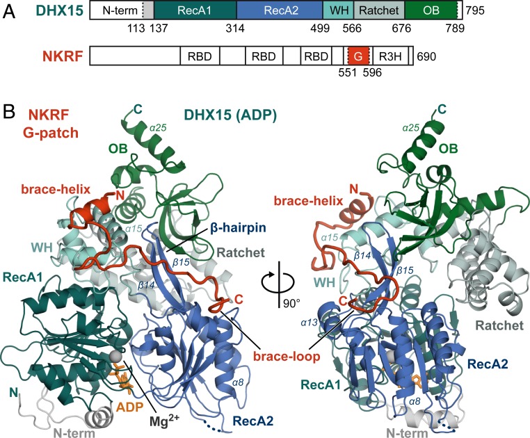
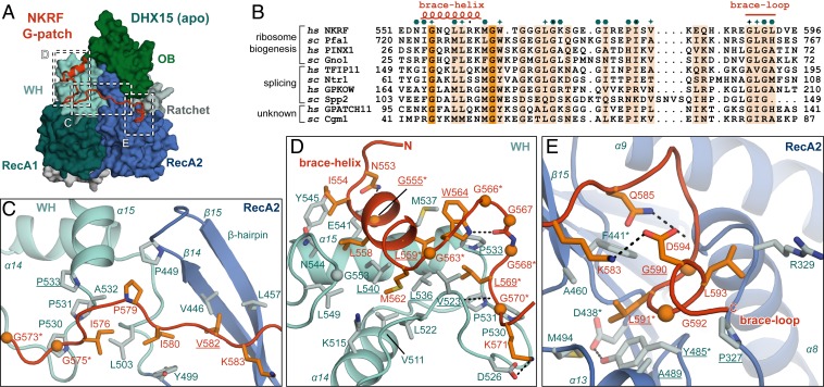
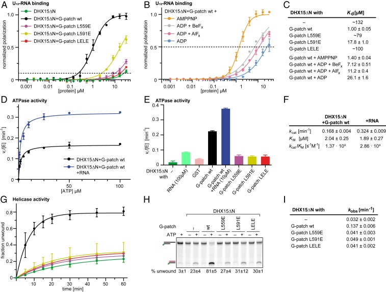
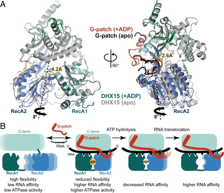
RNA helicases of the DEAH/RHA family are involved in many essential cellular processes, such as splicing or ribosome biogenesis, where they remodel large RNA-protein complexes to facilitate transitions to the next intermediate. DEAH helicases couple adenosine triphosphate (ATP) hydrolysis to conformational changes of their catalytic core. This movement results in translocation along RNA, which is held in place by auxiliary C-terminal domains. The activity of DEAH proteins is strongly enhanced by the large and diverse class of G-patch activators. Despite their central roles in RNA metabolism, insight into the molecular basis of G-patch-mediated helicase activation is missing. Here, we have solved the structure of human helicase DHX15/Prp43, which has a dual role in splicing and ribosome assembly, in complex with the G-patch motif of the ribosome biogenesis factor NKRF. The G-patch motif binds in an extended conformation across the helicase surface. It tethers the catalytic core to the flexibly attached C-terminal domains, thereby fixing a conformation that is compatible with RNA binding. Structures in the presence or absence of adenosine diphosphate (ADP) suggest that motions of the catalytic core, which are required for ATP binding, are still permitted. Concomitantly, RNA affinity, helicase, and ATPase activity of DHX15 are increased when G-patch is bound. Mutations that detach one end of the tether but maintain overall binding severely impair this enhancement. Collectively, our data suggest that the G-patch motif acts like a flexible brace between dynamic portions of DHX15 that restricts excessive domain motions but maintains sufficient flexibility for catalysis.

Keywords: DEAH/RHA helicase; G-patch proteins; ribosome biogenesis; splicing.


Conflict of interest statement

The authors declare no competing interest.

Figures

Fig. 1.
Fig. 1.
The NKRF G-patch tethers two domains of DHX15 together. (A) Domain organization of hsDHX15 and hsNKRF, with domains contained in the expressed constructs highlighted in color. The positions of RecA1, RecA2, WH, Ratchet, and OB domains in DHX15 are shown. In addition to the G-patch motif, NKRF has three RNA binding domains (RBD) and another nucleic acid binding domain termed R3H. Borders of expressed constructs are indicated by dotted lines. (B) Overview of the DHX15–G-patch complex. Domains are colored as in A, and important secondary structure elements described in the text are labeled. The magnesium ion and ADP molecule in the ATPase active site are shown in gray and orange, respectively. An unmodeled loop is indicated by a dotted line.
Fig. 2.
Fig. 2.
Conserved G-patch residues are crucial for forming hydrophobic interactions. (A) Overview of the complex with three different areas of G-patch interaction with DHX15 indicated by dotted frames, which are displayed in detail in CE. Position D corresponds to G-patch site 1, while position E indicates G-patch site 2, and C marks the intervening linker. (B) Sequence alignment of G-patch proteins from yeast and human that have been assigned as activators of DHX15/Prp43 or DHX16/Prp2. Respective hs and sc homologs are grouped, and the corresponding cellular process is indicated. Brace-helix and brace-loop positions as found in NKRF are labeled on top of the alignment. Dark and light orange highlight residues that are identical and more than 70% similar, respectively. Petrol hexagons mark amino acids involved in DHX15 interactions, while stars mark mutated residues. (C) Interface of the G-patch linker region crossing over to RecA2 via the β-hairpin. (D) Interactions of the N-terminal G-patch brace-helix (red/orange) on the WH domain (cyan/light blue). Names of residues mutated in this study are underlined, and residues mutated in previous studies are marked by asterisks. Hydrogen bonds are indicated by black dotted lines. (E) Binding of the C-terminal G-patch brace-loop to the RecA2 domain.
Fig. 3.
Fig. 3.
Mutations in the two binding sites have different effects on complex formation. (AC) Coomassie-stained gels of copurification assays of MBP–DHX15ΔN and GST–NKRF G-patch coexpressed in E. coli. Input (0.03%) and eluates (4.4%) were loaded. GST served as a control. DHX15 expression was verified by Western blotting using an antibody against DHX15. The asterisk marks an unspecific band that copurifies with GST from E. coli lysates in all conditions. DHX15 mutations in G-patch sites 1 and 2 are shown in A and B, respectively. YELE corresponds to double mutant Y485E, L536E, while AELE stands for A489E, L540E. Mutations in the G-patch motif are shown in C. (D) Western blots of coprecipitation assays from HEK293T cells overexpressing HA-2S-NKRF or HA-2S-GFP as a control. Samples were treated with RNase A and tested for copurification of endogenous DHX15. Tubulin served as a loading control. For anti-HA blots, 0.75% of input and 5% of eluates were loaded, while 0.25% of input and 15% of eluates were loaded for anti-DHX15 and anti–α-tubulin blots.
Fig. 4.
Fig. 4.
G-patch binding stabilizes a DHX15 conformation consistent with RNA binding. (A and C) Different conformations of ctPrp43 recorded in the presence (A) or absence (C) of RNA are shown [PDB ID codes 5lta and 5ltk (17)]. The location of G-patch sites 1 and 2 on Prp43 according to superpositions with the DHX15-G-patch complex are indicated by dashed circles. RNA and ADP present in A are shown in black. The location of the RNA binding channel as displayed in A is indicated in C by dashed lines. Rotation of the RecA domains necessary to transform the RNA-bound structure into the RNA-free structure is indicated by an arrow. (B and D) Superpositions of the DHX15–G-patch complex with the structures shown in A and C, respectively.
Fig. 5.
Fig. 5.
NKRF G-patch–mediated enhancement of DHX15 RNA affinity and ATPase activity depends on efficient domain tethering. (A) Fluorescence polarization experiments of FAM-U12 RNA with His10–DHX15ΔN in the absence or presence of GST–NKRF G-patch wt or mutants. The dashed line indicates 10% normalized polarization. Average values of triplicate measurements were plotted, and error bars correspond to SDs. (B) Fluorescence polarization experiments with DHX15ΔN and GST-NKRF G-patch wt in the presence of different ATP analogs or ADP (100 µM). The dashed line indicates 50% normalized polarization. Average values of triplicate measurements were plotted, and error bars correspond to SD. (C) RNA dissociation constants (Kd) with SEM determined by fitting curves in A and B by linear regression. For curves that do not reach 50% normalized polarization, the fitted constants were rounded to two digits and listed as approximate values. (D and F) Michaelis–Menten plot of ATPase activity of His10–DHX15ΔN in complex with GST–NKRF G-patch wt with (blue) or without (black) RNA. Measurements were carried out in triplicate, and error bars represent SD. Michaelis–Menten parameters and corresponding SEM were determined by linear regression. (E) Initial ATP hydrolysis rates at 2 mM ATP, normalized for enzyme concentrations for His10–DHX15ΔN with or without GST–NKRF G-patch wt or mutants, or GST as a control. Error bars indicate SD of triplicate measurements. (G and I) RNA duplex unwinding assays with His10–DHX15ΔN in the presence or absence of GST–NKRF G-patch wt or mutants using FAM-labeled dsRNA with a 3′ single-stranded overhang. Time courses were measured in triplicate, and error bars indicate SD. Rate constants and respective SEM were determined by linear regression. (H) Native PAGE of representative samples from the unwinding assay after 60-min reaction time detecting FAM-fluorescence signal. Samples without ATP or without protein were used as controls for RNase contamination. The top bands correspond to duplex RNA substrate, while the lower bands stem from unwound RNA that is quenched with a cDNA oligo.
Fig. 6.
Fig. 6.
G-patch binding retains RecA2 motions in DHX15 induced by ADP binding. (A) Superposition of the DHX15–G-patch complex without (apo; gray/black) or with ADP bound in the active site (blue-green/red). The RecA2 domain rotation required to transform the apo into the ADP-bound structure is indicated by a thick black arrow, while the imaginary rotation axis through the back of RecA2 is shown as a dashed line. Shifts in RecA2 and the G-patch brace-loop are indicated. Domain movements were analyzed by using the Dyndom server (79). (B) Suggested model for G-patch–mediated stimulation of DEAH RNA-helicases. The C-terminal domains including the WH are depicted in cyan, and RecA1 and RecA2 are colored in green and blue, respectively. Mobility is indicated by multiple lighter-shaded schematics.

References

    1. Bleichert F., Baserga S. J., The long unwinding road of RNA helicases. Mol. Cell 27, 339–352 (2007). - PubMed
    1. Bourgeois C. F., Mortreux F., Auboeuf D., The multiple functions of RNA helicases as drivers and regulators of gene expression. Nat. Rev. Mol. Cell Biol. 17, 426–438 (2016). - PubMed
    1. Cordin O., Beggs J. D., RNA helicases in splicing. RNA Biol. 10, 83–95 (2013). - PMC - PubMed
    1. Jarmoskaite I., Russell R., RNA helicase proteins as chaperones and remodelers. Annu. Rev. Biochem. 83, 697–725 (2014). - PMC - PubMed
    1. Ozgur S., et al. , The conformational plasticity of eukaryotic RNA-dependent ATPases. FEBS J. 282, 850–863 (2015). - PubMed

Publication types

LinkOut - more resources