. 2020 Dec 25;295(52):18379-18389.
doi: 10.1074/jbc.RA120.012395. Epub 2020 Oct 29.

A highly potent CD73 biparatopic antibody blocks organization of the enzyme active site through dual mechanisms

Affiliations

A highly potent CD73 biparatopic antibody blocks organization of the enzyme active site through dual mechanisms

James E Stefano et al. J Biol Chem. .

Abstract

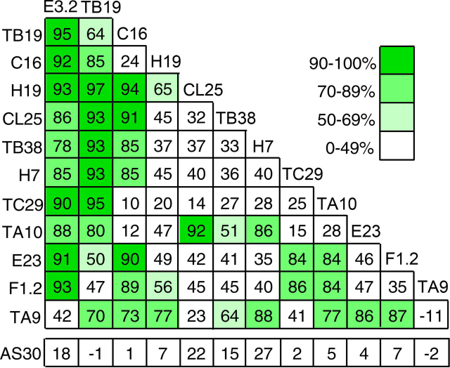
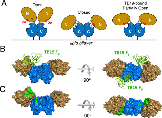
The dimeric ectonucleotidase CD73 catalyzes the hydrolysis of AMP at the cell surface to form adenosine, a potent suppressor of the immune response. Blocking CD73 activity in the tumor microenvironment can have a beneficial effect on tumor eradication and is a promising approach for cancer therapy. Biparatopic antibodies binding different regions of CD73 may be a means to antagonize its enzymatic activity. A panel of biparatopic antibodies representing the pairwise combination of 11 parental monoclonal antibodies against CD73 was generated by Fab-arm exchange. Nine variants vastly exceeded the potency of their parental antibodies with ≥90% inhibition of activity and subnanomolar EC50 values. Pairing the Fabs of parents with nonoverlapping epitopes was both sufficient and necessary whereas monovalent antibodies were poor inhibitors. Some parental antibodies yielded potent biparatopics with multiple partners, one of which (TB19) producing the most potent. The structure of the TB19 Fab with CD73 reveals that it blocks alignment of the N- and C-terminal CD73 domains necessary for catalysis. A separate structure of CD73 with a Fab (TB38) which complements TB19 in a particularly potent biparatopic shows its binding to a nonoverlapping site on the CD73 N-terminal domain. Structural modeling demonstrates a TB19/TB38 biparatopic antibody would be unable to bind the CD73 dimer in a bivalent manner, implicating crosslinking of separate CD73 dimers in its mechanism of action. This ability of a biparatopic antibody to both crosslink CD73 dimers and fix them in an inactive conformation thus represents a highly effective mechanism for the inhibition of CD73 activity.

Keywords: CD73; aggregation; antibody engineering; enzyme mechanism; enzyme structure; protein complex; protein crosslinking; structure-function.


Conflict of interest statement

Conflict of interest—All authors are or were employees of Sanofi.

Figures

Figure 1
Figure 1
Screen of inhibitory activity against CD73 on COR-L23 cells. The inhibition of CD73 activity (%) was determined following exposure to antibodies for 4 h using a LC-MS–based assay with a heavy-isotope AMP substrate (white shading: 0–49% inhibition at 1 ug/ml; pale green: 50–69% inhibition; green: 70–89% inhibition; and dark green: 90–100% inhibition). Each square, except the furthest right in each row, represents a biparatopic produced by the combination of parental antibodies indicated on the horizontal and vertical axes. The furthest right square in each row represents the parental bivalent anti-CD73 antibody reconstructed using Fab-arm exchange. The bottom row (AS30) indicates pairings with an irrelevant antibody AS30, to produce monovalent versions of the parental antibodies. Values represent the mean inhibition at 1 μg/ml observed with four replicates of single analyte dilutions.
Figure 2
Figure 2
Relative affinities of parental and biparatopic antibodies for CD73. Each parental antibody in monovalent form having a second irrelevant arm (AS30) and the biparatopic variant were immobilized and exposed to soluble CD73 in the flow. KD values are based on association rate constants using a 1:1 Langmuir binding model combined with dissociation rate constants based on either a 1:1 Langmuir model or the principal more stable, component of dissociation where biphasic dissociation kinetics were observed. Raw sensorgrams can be found in Fig. S8, S9.
Figure 3
Figure 3
Epitope binning by biolayer interferometry (Octet). A mixture of CD73 with a molar excess of Fab was incubated with monovalent parental antibodies immobilized on a solid support. A, cartoon of the assay format showing the condition of nonoverlapping epitopes and no blocking (top panel) or overlapping epitopes producing complete blocking of capture (bottom panel). B, capture of CD73/Fab complexes by immobilized antibodies. Capture was normalized to the signal from CD73 alone in the absence of Fab as described under “Experimental Procedures.” Raw Octet traces can be found in Fig. S9. C, epitope binning based on the inhibition of capture. D, inhibition of CD73 activity on COR-L23 cells versus the ability of antibodies to capture a CD73/Fab complex in vitro. Gray-filled circles: capture of a CD73/Fab complex of the same antibody on the support (parental pair).
Figure 4
Figure 4
Structure of TB19 with CD73.A, cartoon representation of the different conformational states of CD73. The CD73 N-terminal domain of CD73 is shown in beige and the C-terminal domain is shown in blue. Gray coils: linker elements connecting the two domains. Red spheres: zinc cofactor(s) bound by the N-terminal domain. S: substrate. Note that the structure of the complex with TB19 was obtained in the absence of substrate, which is not shown. B, two TB19 Fv domains binding one CD73 dimer in the intermediate conformation viewed from two different angles. Colors are as in A with the zinc and phosphate molecules shown as red spheres. The TB19 Fab (green) is shown in a cartoon representation. C, mapping of the epitope for TB19 on CD73. The coloring scheme is as B; residues interacting withTB19 are shown in green.
Figure 5
Figure 5
Structures of TB38 with CD73. CD73 coloring scheme: beige = N-terminal domain, blue = C-terminal domain, gray = linker. A, TB38 Fab::CD73 structure with CD73 in the open conformation (chains A and B in PDB ID 6VCA). Numbers identify each monomer TB38 Fab is shown in a purple cartoon representation. B, TB38 Fv::CD73 structure with the CD73 in an open/closed hybrid conformation (chains C and D in PDB 6VCA). Monomer 1 is in the open conformation and monomer 2 in the closed conformation (see text). TB38 Fv is shown in purple. C, mapping of the TB38 epitope residues (purple) on CD73 in the open/closed hybrid conformation as shown in (B).
Figure 6
Figure 6
Potential modes of co-engagement of CD73 by the TB19/TB38 biparatopic. Bispecific antibodies are modeled based on TB19:CD73, TB38:CD73, and full-IgG1 (PDB 1ZHZ) structures. A, surface representation of a TB19/TB38 biparatopic antibody. Arrow: distance between the last residues in the CH1 domains. The TB19 Fab is shown in green, the TB38 Fab in purple. The Fc is shown light gray. B, a model of four TB19/TB38 biparatopic antibodies bound by a CD73 dimer in the partially open configuration, as seen for the complex with TB19. The N-terminal domains of CD73 are shown in beige and the C-terminal domains in blue. Arrow: distances separating the last residues of the CH1 domains of the opposing Fabs in adjacent biparatopic antibodies.

References

    1. Allard D., Allard B., Gaudreau P.O., Chrobak P., Stagg J. CD73-adenosine: A next-generation target in immuno-oncology. Immunotherapy. 2016;8:145–163. doi: 10.2217/imt.15.106. 26808918. - DOI - PubMed
    1. Allard B., Longhi M.S., Robson S.C., Stagg J. The ectonucleotidases CD39 and CD73: Novel checkpoint inhibitor targets. Immunol. Rev. 2017;276:121–144. doi: 10.1111/imr.12528. 28258700. - DOI - PMC - PubMed
    1. Allard D., Chrobak P., Allard B., Messaoudi N., Stagg J. Targeting the CD73-adenosine axis in immuno-oncology. Immunol. Lett. 2019;205:31–39. doi: 10.1016/j.imlet.2018.05.001. 29758241. - DOI - PubMed
    1. Kats L., Darcy P.K., Beavis P.A., Bahreyni A., Rezaei M., Khazaei M., Fuiji H., Ferns G.A., Ryzhikov M., Avan A., Hassanian S.M. The potential role of adenosine signaling in the pathogenesis of melanoma. Int. J. Mol. Sci. 2018;156:451–457. doi: 10.1016/j.bcp.2018.09.018. 30232037. - DOI - PubMed
    1. Sek K., Mølck C., Stewart G.D. Targeting adenosine receptor signaling in cancer immunotherapy. 2018;19 doi: 10.3390/ijms19123837. 30513816. - DOI - PMC - PubMed

MeSH terms