. 2024 Sep 11;15(1):7969.
doi: 10.1038/s41467-024-52251-3.

Phalloidin and DNase I-bound F-actin pointed end structures reveal principles of filament stabilization and disassembly

Affiliations

Phalloidin and DNase I-bound F-actin pointed end structures reveal principles of filament stabilization and disassembly

Micaela Boiero Sanders et al. Nat Commun. .

Abstract

Actin filament turnover involves subunits binding to and dissociating from the filament ends, with the pointed end being the primary site of filament disassembly. Several molecules modulate filament turnover, but the underlying mechanisms remain incompletely understood. Here, we present three cryo-EM structures of the F-actin pointed end in the presence and absence of phalloidin or DNase I. The two terminal subunits at the undecorated pointed end adopt a twisted conformation. Phalloidin can still bind and bridge these subunits, inducing a conformational shift to a flattened, F-actin-like state. This explains how phalloidin prevents depolymerization at the pointed end. Interestingly, two DNase I molecules simultaneously bind to the phalloidin-stabilized pointed end. In the absence of phalloidin, DNase I binding would disrupt the terminal actin subunit packing, resulting in filament disassembly. Our findings uncover molecular principles of pointed end regulation and provide structural insights into the kinetic asymmetry between the actin filament ends.


Conflict of interest statement

The authors declare no competing interests.

Figures

Fig. 1
Fig. 1. Phalloidin bridges and flattens both terminal actin subunits at the pointed end.
a Exemplary 2D classes of the undecorated pointed end of actin filaments. Box size: 324 × 324 Å. b Cryo-EM density map of the undecorated pointed end of F-actin at a resolution of 3.1 Å. Actin subunits are labeled depending on their location on the filament, so that the ultimate subunit is P0 and subunits towards the filament center have an increasing number (P1, P2, P3). The ultimate (P0) and penultimate (P1) actin subunits are colored in purple and lilac, respectively, while the subunits towards the filament center (P2 and beyond) are colored in light grey. The bound nucleotide is highlighted in yellow. c Cryo-EM density map of the phalloidin-bound pointed end of F-actin at a resolution of 3.7 Å. Phalloidin-bound actin subunits are colored from dark orange at the pointed end to lighter orange towards the filament center. The bound nucleotide and phalloidin are colored yellow and cyan, respectively. d Superimposition of the last four subunits (P0-P3) of the undecorated pointed end (purple and light grey) and the phalloidin-bound pointed end (shades of orange). The four actin subdomains (SD1-4), nucleotide binding site and hydrophobic cleft are annotated. The black arrow highlights the ~12° rotation of the outer subdomains (SD1, SD2) relative to the inner subdomains (SD3, SD4).
Fig. 2
Fig. 2. Binding site of phalloidin to the ultimate and penultimate actin subunits at the pointed end.
a Structure of the phalloidin-bound pointed end. Actin subunits are labelled from P0 (ultimate actin subunit at the pointed end) to P2 (actin subunit towards the filament center). Phalloidin-bound actin subunits are colored from dark orange at the pointed end to lighter orange towards the filament center. The bound nucleotide and phalloidin are colored yellow and cyan, respectively. b Zoom-in on the phalloidin binding site at the pointed end. The amino acids from subunits P0 and P1 that interact with phalloidin are annotated and shown with stick representation. c Clipped surface representation of the ultimate phalloidin binding site. d Structure of the undecorated pointed end. The ultimate (P0) and penultimate (P1) actin subunits are colored in purple and lilac, respectively, while the subunits towards the filament center (P2 and beyond) are colored in light grey. The bound nucleotide is highlighted in yellow. e Zoom-in on the interface between P0 and P1, superimposing the phalloidin-bound (orange) and undecorated (purple) pointed end. The phalloidin-induced displacement of various actin loops is indicated. f Surface representation of the undecorated pointed end with a fitted phalloidin in the ultimate phalloidin binding site. Phalloidin at this position would clash with actin.
Fig. 3
Fig. 3. Two DNase I molecules bind to the pointed end of actin filaments.
a Exemplary 2D classes of the DNase I-bound pointed-end of actin filaments (conformer 1), stabilized with phalloidin. Light green arrowheads mark the position of the two DNase I molecules. Box size: 282 × 282 Å. b Cryo-EM density map of the DNase I-bound pointed end of F-actin at a resolution of 3.8 Å (conformer 1). Actin subunits are labelled from P0 (ultimate actin subunit at the pointed end) to P2 (actin subunit towards the filament center). Phalloidin-bound actin subunits are colored from dark orange at the pointed end to lighter orange towards the filament center. The two bound DNase I molecules, the bound nucleotide and phalloidin are colored light green, yellow and cyan, respectively. c Structure of the DNase I-bound pointed end of F-actin corresponding to conformer 1 (left). Structure of conformers 1 and 2 (modeled after maps obtained by 3D classification), see also the 3D variability results in Supplementary Movie 4 (middle and right). d, e Zoom-in on the two major interfaces between the D-loop of actin and DNase I (conformer 1).
Fig. 4
Fig. 4. DNase I bound to the penultimate actin subunit (P1) displaces the ultimate actin subunit (P0).
a Structure of the undecorated pointed end (left, purple and grey) and the penultimate subunit P1 of the phalloidin-bound pointed end bound by DNase I (middle, orange and light green) highlighting subdomain 2 (SD2). The SD2s were used for the alignment of the two structures, yielding a model of the native pointed end decorated by DNase I (right). Actin subunits are labeled depending on their location on the filament, so that the ultimate subunit is P0 and subunits towards the filament center have an increasing number (P1, P2, P3). The bound nucleotide is highlighted in yellow. b Zoom-in image of the clashes between the penultimate DNase I and the actin subunit P0. The clashing region is shown in pink.
Fig. 5
Fig. 5. Model of DNase I-mediated disassembly of actin filaments.
The model depicts the pointed end of an actin filament, with the flattened subunits in grey and the twisted subunits in shades of purple, the ultimate subunit is labeled P0 and the subsequential actin subunits towards the filament center are labeled with increasing numbers (P1-P4). A DNase I molecule (depicted in light green) can bind to each of the last two actin subunits of the pointed end. DNase I binding to the penultimate subunit displaces the ultimate actin subunit from the pointed end, releasing a G-actin molecule (depicted in pink) bound to DNase I. For more a detail explanation we refer to the Results section.
Fig. 6
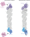
Fig. 6. Model explaining asymmetry in the incorporation and loss of subunits at opposite filament ends.
Actin monomers are colored in pink. The internal subunits of actin filaments are colored in grey, whereas the last two subunits of the barbed end and the pointed end are colored in shades of blue and purple, respectively. Subunits of the pointed end are labeled Px, with x starting from 0 with increasing numbers towards the filament center, and subunits at the barbed end are labeled in a similar manner with Bx. Subunits labeled G are in a twisted, G-actin like conformation, and subunits labeled F are in a flattened, filament-like conformation. Subunits labeled G to F and F to G are those that are expected to undergo a conformational change during polymerization and depolymerization, respectively. For a detailed discussion of this model we refer to the Discussion section.

References

    1. Pollard, T. D. & Cooper, J. A. Actin, a Central Player in Cell Shape and Movement. Science326, 1208–1212 (2009). 10.1126/science.1175862 - DOI - PMC - PubMed
    1. Blanchoin, L., Boujemaa-Paterski, R., Sykes, C. & Plastino, J. Actin Dynamics, Architecture, and Mechanics in Cell Motility. Physiol. Rev.94, 235–263 (2014). 10.1152/physrev.00018.2013 - DOI - PubMed
    1. Kabsch, W., Mannherz, H. G., Suck, D., Pai, E. F. & Holmes, K. C. Atomic structure of the actin: DNase I complex. Nature347, 37–44 (1990). 10.1038/347037a0 - DOI - PubMed
    1. Oda, T., Iwasa, M., Aihara, T., Maéda, Y. & Narita, A. The nature of the globular- to fibrous-actin transition. Nature457, 441–445 (2009). 10.1038/nature07685 - DOI - PubMed
    1. Dominguez, R. & Holmes, K. C. Actin Structure and Function. Annu. Rev. Biophys.40, 169–186 (2011). 10.1146/annurev-biophys-042910-155359 - DOI - PMC - PubMed

Publication types

LinkOut - more resources