. 2024 Nov 20;15(1):10038.
doi: 10.1038/s41467-024-54212-2.

The inhibitory action of the chaperone BRICHOS against the α-Synuclein secondary nucleation pathway

Affiliations

The inhibitory action of the chaperone BRICHOS against the α-Synuclein secondary nucleation pathway

Dhiman Ghosh et al. Nat Commun. .

Abstract

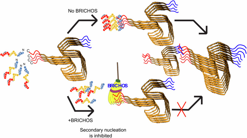
The complex kinetics of disease-related amyloid aggregation of proteins such as α-Synuclein (α-Syn) in Parkinson's disease and Aβ42 in Alzheimer's disease include primary nucleation, amyloid fibril elongation and secondary nucleation. The latter can be a key accelerator of the aggregation process. It has been demonstrated that the chaperone domain BRICHOS can interfere with the secondary nucleation process of Aβ42. Here, we explore the mechanism of secondary nucleation inhibition of the BRICHOS domain of the lung surfactant protein (proSP-C) against α-Syn aggregation and amyloid formation. We determine the 3D NMR structure of an inactive trimer of proSP-C BRICHOS and its active monomer using a designed mutant. Furthermore, the interaction between the proSP-C BRICHOS chaperone and a substrate peptide has been studied. NMR-based interaction studies of proSP-C BRICHOS with α-Syn fibrils show that proSP-C BRICHOS binds to the C-terminal flexible fuzzy coat of the fibrils, which is the secondary nucleation site on the fibrils. Super-resolution fluorescence microscopy demonstrates that proSP-C BRICHOS runs along the fibrillar axis diffusion-dependently sweeping off monomeric α-Syn from the fibrils. The observed mechanism explains how a weakly binding chaperone can inhibit the α-Syn secondary nucleation pathway via avidity where a single proSP-C BRICHOS molecule is sufficient against up to ~7-40 α-Syn molecules embedded within the fibrils.


Conflict of interest statement

Competing interests: The authors declare no competing interest.

Figures

Fig. 1
Fig. 1. NMR Structure of the proSP-C BRICHOS domain.
a X-ray crystallography structure (2YAD) of the proSP-C BRICHOS domain in a ribbon representation demonstrating the existence of a homotrimer (with the individual entities colored red, green and blue) and b the corresponding NMR structure represented by 20 conformers with the long loop forming residues Gln151-Phe180, which are absent in the crystal structure, are highlighted by lighter colors. c Superposition of the NMR structure of the proSP-C BRICHOS domain colored in dark green and light green (loop) with the x-ray crystallography structure (2YAD) in red.
Fig. 2
Fig. 2. NMR structure of monomeric proSP-C BRICHOS variant II.
a Salt bridge mutation sites of trimeric WT proSP-C BRICHOS that are perturbed in the Var II to generate a stable monomeric form. b The solution state NMR structure of the proSP-C BRICHOS mutant (Var II) shows formation of a monomer in contrast to the trimer formed by WT proSP-C BRICHOS. c Monomeric proSP-C BRICHOS exhibits an extended hydrophobic cleft as highlighted by yellow color, which is believed to be the active site for the chaperone action. d Structure overlay of one monomer from the WT proSP-C BRICHOS trimer (green) and the monomer of the proSP-C BRICHOS mutant (Var II) (cyan for β-sheet) showing that secondary structure elements are conserved in the monomeric proSP-C BRICHOS Var II despite repositioning of the loop.
Fig. 3
Fig. 3. Binding site of a client peptide on the pro-SP-C BRICHOS domain.
a [15N,1H]-TROSY spectra of 15N-labeled proSP-C BRICHOS Var II in absence (red) and presence of 200 µM (green),500 µM (yellow), 1 mM (cyan) and 2 mM (blue) of client peptide B. b Client peptide B-induced chemical shift perturbations (CSPs) versus the amino acid sequence of proSP-C BRICHOS Var II with secondary structure elements indicated. c Binding sites for interaction of client peptide B mapped on the proSP-C BRICHOS Var II domain. Residues with CSPs > 0.03 ppm are shown in green color.
Fig. 4
Fig. 4. Monomeric proSP-C BRICHOS Var II inhibits Aβ42 fibrillation more efficiently than WT.
a Aggregation of Aβ42 in the presence of different proSP-C BRICHOS variants. Aggregation of Aβ42 in presence of varying molar equivalent of (left panel) WT, Var I (middle panel), and Var II (right panel). The increase in fibrillar mass on the y-axis was measured as an increase in the fluorescence of thioflavin T (ThT). The points represent individual data points. The solid lines represent the fits as obtained from amylo fit as described by Meisl et al. b Binding curves for the interaction between different proSP-C BRICHOS variants and Aβ42 fibrils. Left panel: Binding curve for the interaction between Aβ42 fibrils and WT BRICHOS at three different BRICHOS concentrations 300 nM (red), 600 nM (magenta), 900 nM (blue), yielding a dissociation constant, Kd ~ 191.7 [6.2; 517.0] nM with a stoichiometry of 1 BRICHOS molecule per ~8 [5; 12] monomer units in the fibril. Middle panel: Binding curve for the interaction between Aβ42 fibrils and proSP-C BRICHOS variant I at three different BRICHOS concentrations, 250 nM (red), 375 nM (magenta) and 500 nM (blue). The data show no significant binding. Right panel: Binding curve for the interaction between Aβ42 fibrils and proSP-C BRICHOS variant II at three different BRICHOS concentrations, 150 nM (red), 300 nM BRICHOS (magenta), and 600 nM (blue). This data yields a dissociation constant, Kd ~ 21.4 [0.1; 290.4] nM with a stoichiometry of 1 BRICHOS molecule per ~6 [2; 9] monomer units in the fibril. Each experiment was performed 3 times. Error bars are derived from the standard deviation and corresponding mean +/- S.D are represented here.
Fig. 5
Fig. 5. Influence of different proSP-C BRICHOS variants on binding to α-Syn fibrils and α-Syn fibrillation pathway.
a Aggregation of α-Syn in presence of different proSP-C BRICHOS variants. Aggregation of α-Syn in presence of varying molar equivalents of WT (left panel), Var I (middle panel) and Var II (right panel). The increase in fibrillar mass was measured as an increase in the fluorescence of thioavin T (ThT). The points represent individual data points, the solid lines represent the fits as obtained from amylo fit (Meisl et al.). b Binding curves for the interaction between different proSP-C BRICHOS variants and α-Syn fibrils. (left panel) Binding curve for the interaction between α-Syn fibrils and WT proSP-C BRICHOS at three different concentrations: 150 nM (red), 300 nM (magenta), 600 nM (blue). This yielded a dissociation constant, Kd = Kd  = ~695.7 [263.1, 1311] nM with a stoichiometry of 1 BRICHOS molecule per ~38 [22; 56] monomer units in the fibrils. (Middle panel) Binding curve for the interaction between α-Syn fibrils and proSP-C BRICHOS Var I at three different concentrations, 150 nM (red), 300 nM (magenta), and 600 nM (blue). This yielded a dissociation constant, Kd  ~ 1.26 [0.09; 2.96] nM with a stoichiometry of 1 BRICHOS molecule per ~~18 [12; 60] monomer units in the fibril. (Right panel) Binding curve for the interaction between α-Syn fibrils and proSP-C BRICHOS variant II at three different BRICHOS concentrations: 300 nM (red), 600 nM (magenta), and 900 nM (blue). This yielded a dissociation constant, Kd ~ 450.2 [109.8, 1005] μM with a stoichiometry of ~7 [4; 10] monomer units in the fibril. Each experiment was performed 3 times. Error bars are derived from the standard deviation and corresponding mean +/- S.D are represented here.
Fig. 6
Fig. 6. ProSP-C BRICHOS interacts with the flexible C-terminal part of α-Syn fibrils, which is the secondary nucleation site.
a Competition experiment on α-Syn fibrils (Syn fib) with 15N-labeled α-Syn monomer (Syn Mono) measured by [15N,1H]-HMQC experiments against the addition of proSP-C BRICHOS (BRI) Var II. The [15N,1H]-HMQC of 15N-labeled α-Syn monomer only (orange, left) is the reference spectrum yielding the I0 values for panel b, shown along-side [15N,1H]-HMQC spectra of α-Syn monomer in absence (red, middle) and presence (blue, right) of proSP-C BRICHOS Var II while bound to α-Syn fibrils. b Intensity ratios (I/I0) relative to the control measurement with free α-Syn of individual backbone15N-1H moieties of monomeric α-Syn in presence of α-Syn fibrils (red) or α-Syn fibrils and proSP-C BRICHOS Var II (blue). Signal loss is observed due to the transient binding of monomeric α-Syn to its fibrils (red). Upon addition of proSP-C BRICHOS Var II, signal loss is attenuated, which can be attributed to a competitive binding between proSP-C BRICHOS and monomeric α-Syn on the fibrils. c Monomeric α-Syn release attached to α-Syn fibrils upon addition of proSP-C BRICHOS Var II. The experimental set up is as follows: In a sample with 540 µM unlabeled α-Syn amyloid fibrils 100 μM 15N-labeled monomeric α-Syn is added and incubated for two hours. Next, 100 μM (1:1) or 300 μM (1:3) or 500 μM (1:5) proSP-C BRICHOS Var II were added to the sample at time point 0 and the intensity of the 15N-labeled monomeric α-Syn is measured time-resolved by a 15N-filtered NMR experiment (i.e. [15N,1H]-HMQC) yielding after ca 120 min ~ 25% (for 1:1), ~50% (for 1:3) and ~65% (1:5) monomer bound to fibrils were released as plotted in bar diagram (d). e [15N,1H]-HMQC spectra of 15N-labeled proSP-C BRICHOS Var II in absence and in presence of WT α-Syn fibrils (blue and cyan, respectively) or in presence of α-Syn(1-121) fibrils (red). Signal attenuation of free proSP-C BRICHOS Var II is observed indicating its binding to the fibrils. f Overall intensity ratios (I/I0) of the signals in the [15N, 1H]-HMQC spectra of proSP-C BRICHOS Var II (blue)in presence of WT α-Syn (cyan) or α-Syn(1-121) fibrils (red). I0 corresponds to free proSP-C BRICHOS Var II in buffer, while I correspond to its respective state in presence of the fibrils.
Fig. 7
Fig. 7. Single particle tracking microscopy of ProSP-C BRICHOS Var II shows 1D diffusion along α-Syn fibrils.
a Single tracks of proSP-C BRICHOS Var II at 10 nM are overlaid with a TIRF microscopy frame showing all trajectories that are longer than 10 frames. b Diffusion coefficients are calculated for at least 1000 trajectories and filtered to exclude static molecules (D > 0.05 µm2/s). Error bars represent standard deviation. The insets depict 3 examples of long trajectories (color code represents a time of 100 seconds). c The mean square displacement for all trajectories is plotted versus the time lag. The first 3 data points are taken to estimate the slope. At higher concentrations (10 and 100 nM) the slope at high time lags, suggesting a sub diffusional character. The insets are six representative isolated tracks along α-Syn fibrils from a. For 1nM, 10nM and 100nM no of data points are collected 8545, 2445 and 757, respectively. Error bars are derived from the standard deviation and corresponding mean +/- S.D are represented here.
Fig. 8
Fig. 8. Schematic representation of the secondary nucleation inhibition of α-Syn aggregation and fibrillation by the chaperone proSP-C BRICHOS.
α-Syn monomers converted into fibrils during its aggregation pathway. Monomeric synuclein binds to the C-terminal flexible tail of the fibrils allowing acceleration of the aggregation process via secondary nucleation. ProSP-C BRICHOS competes with the same binding site on the fibrils, preventing further binding of monomers and cleaning the existing monomers that are already bound to the fibrils.

References

    1. Spillantini, M. G. et al. Alpha-synuclein in Lewy bodies. Nature388, 839–40 (1997). - PubMed
    1. Lashuel, H. A., Overk, C. R., Oueslati, A. & Masliah, E. The many faces of α-synuclein: from structure and toxicity to therapeutic target. Nat. Rev. Neurosci.14, 38–48 (2013). - PMC - PubMed
    1. Prymaczok, N. C., Riek, R. & Gerez, J. More than a Rumor Spreads in Parkinson’s Disease. Front Hum. Neurosci.10, 608 (2016). - PMC - PubMed
    1. Krüger, R. et al. Ala30Pro mutation in the gene encoding alpha-synuclein in Parkinson’s disease. Nat. Genet18, 106–8 (1998). - PubMed
    1. Ibáñez, P. et al. Causal relation between alpha-synuclein gene duplication and familial Parkinson’s disease. Lancet364, 1169–71 (2004). - PubMed

Publication types

MeSH terms

LinkOut - more resources