. 2024 Oct 9;15(1):8740.
doi: 10.1038/s41467-024-52891-5.

Mycobacterial HelD connects RNA polymerase recycling with transcription initiation

Affiliations

Mycobacterial HelD connects RNA polymerase recycling with transcription initiation

Tomáš Kovaľ et al. Nat Commun. .

Abstract

Mycobacterial HelD is a transcription factor that recycles stalled RNAP by dissociating it from nucleic acids and, if present, from the antibiotic rifampicin. The rescued RNAP, however, must disengage from HelD to participate in subsequent rounds of transcription. The mechanism of release is unknown. We show that HelD from Mycobacterium smegmatis forms a complex with RNAP associated with the primary sigma factor σA and transcription factor RbpA but not CarD. We solve several structures of RNAP-σA-RbpA-HelD without and with promoter DNA. These snapshots capture HelD during transcription initiation, describing mechanistic aspects of HelD release from RNAP and its protective effect against rifampicin. Biochemical evidence supports these findings, defines the role of ATP binding and hydrolysis by HelD in the process, and confirms the rifampicin-protective effect of HelD. Collectively, these results show that when HelD is present during transcription initiation, the process is protected from rifampicin until the last possible moment.


Conflict of interest statement

The authors declare no competing interests.

Figures

Fig. 1
Fig. 1. Associating partners of HelD in vivo and structure of the Msm RNAP core together with σA, RbpA and HelD.
a Silver-stained SDS-PAGE of HelD-FLAG pull-down from exponential (EXP) and stationary (STA) phase of growth. A No-Tag strain was used as a control. Proteins pulled down with HelD are indicated on the right-hand side. The experiment was performed four times and a representative gel is shown. The dotted line shows electronic assembly of the gel. b Quantitative mass spectrometry analysis of HelD-FLAG pull-down vs No-Tag strain in EXP and STA phases of growth, respectively. The analysis was done from three biological replicates. The abundance of individual proteins was compared by two-tailed student’s t-test. The permutation-based FDR was used as an adjustment of p-value. The enrichment is shown with a volcano plot (−log10 p value > 2 on the y-axis, protein enrichments > 1.5 on the x-axis). Significantly enriched proteins are shown as red (EXP) and blue (STA) dots, respectively. The identity of the most enriched proteins is indicated. c Enrichment of selected proteins from (b) (EXP and STA) related to the transcription machinery, showing relative enrichment of the proteins in the HelD-FLAG pull-down. CarD was not present in the HelD-FLAG pull-down dataset. This is indicated with the cross. Source data are provided as a Source Data file. d Color-coded annotation of Msm RNAP core, domains of HelD, σA and RbpA. e, f Two conformations of the Msm RNAP core complex together with σA, RbpA and HelD in state I (HelD-holo-I) and state II (HelD-holo-II), respectively. Individual domains are color-coded according to (d). g Magnified details of panel (f). The mutual interaction of σA and HelD in the context of the β′-clamp. σA2 interacts with the conserved binding site on the β′-clamp coiled-coil domain (β′-clamp CC, gray) near the β′-clamp rudder (green). The σAN-helix and adjacent regions (red) wrap with specific protein–protein interactions around the HelD–CO-tip helix-turn-helix (HTH) motif (light blue). The HelD–CO-tip is also buttressed by helices α1 and α4 of the σA2 domain (purple). h Magnified details of panel (g). Specific residues important for the σA–HelD interaction are highlighted. σA/Phe140 (red) and its interaction with HelD defines the beginning of ordered regions of σA.
Fig. 2
Fig. 2. DNA upstream fork promoter binds to Msm HelD–σA–RbpA–RNAP complex.
a Sequence of the us-fork promoter DNA fragment. The numbers above denote the DNA position with respect to the transcription start site (+1). The −35 and −10 elements are colored yellow, nt/t denotes non-template/template strand, respectively. bd Three conformations of the Msm RNAP core complex together with us-fork promoter DNA fragment, σA, RbpA, and HelD. One conformation is in state II (us-fork-HelD–RPc-II) and two conformations are in state III (us-fork–HelD–RPc-III, us-fork–HelDN-term–RPc-III), respectively. In us-fork–HelD–RPc-II, the whole HelD protein is ordered on RNAP, in us-fork–HelD–RPc-III the 1A–2A NTPase is disengaged and thus HelD–CO tilts relative to the primary channel. In us-fork-HelDN-term-RPc-III, only the HelDN-term domain is bound in the secondary channel and the rest of the HelD protein is not ordered. Individual domains are color-coded as defined in Fig. 1d. eg Close-up views of the RNAP primary channel, corresponding to panels (bd), respectively. The black scale bar illustrates the distance between the β-lobe and the N-terminus of the σA2 domain, which directly correlates with the primary channel closure according to Supplementary Table 3. e Presence of HelD–CO (light blue) keeps the RNAP primary channel wide open. The σAN-helix wraps specifically around the HelD–CO-tip. f Tilting (compare HelD–CO axes) of HelD–CO disfavors CO-tip interaction with the σA2 domain and prevents CO-tip interaction with the σAN-helix. g Displacement of all HelD domains, except for HelDN-term(as depicted in d), allows a partial closure of the RNAP primary channel but not to the extent that would allow the σAN-helix interaction with the β-lobe as seen in the σA–RbpA–RNAP complex (Supplementary Fig. 8d).
Fig. 3
Fig. 3. HelD release on the pathway towards RPo complex formation.
a Sequence of the promoter transcription bubble DNA fragment. The numbers above denote the DNA position with respect to the TSS (+1). The −35 and −10 elements are colored yellow; nt/t denotes non-template/template strand, respectively. b Msm RNAP core complex together with the promoter transcription bubble DNA fragment, σA, RbpA and HelD in the RP2 like-state (HelDN-term–RP2). Only the HelDN-term domain is present in the secondary channel, the rest of the HelD protein is not ordered. The dwDNA is partially loaded into the primary channel. Individual domains are color-coded as in Fig. 1d. c, d Msm RNAP core complex together with the promoter transcription bubble DNA fragment, σA, RbpA, (no HelD), in the RP2-like state (σAN-helix–RP2) and Mtb RP2 RNAP complex (RP2 promoter–DNA–σA–RNAP) PDB 6EE8, respectively. Individual domains are color-coded as in Fig. 1d, CarD in panel d is transparent green. eg Close-up views of the RNAP primary channel from panels (bd), respectively. The black scale bar illustrates the distance between the β-lobe and the N-terminus of the σA2 domain, which directly correlates with the primary channel closure according to Supplementary Table 3. e The presence of HelDN-term (firebrick) in the secondary channel prevents the RNAP primary channel from closing completely. Concomitantly, dwDNA is only partially loaded into the primary channel. f Displacement of the HelDN-term domain is followed by a slight adjustment of the RNAP primary channel and interaction of σAN-helix with the dwDNA. g In the RP2 complex (PDB 6EE8), the RNAP primary channel closes around dwDNA so that the σAN-helix directly interacts with the β-lobe domain. CarD interacts with the −10 element and stabilizes the transcription bubble.
Fig. 4
Fig. 4. NTPase activities of HelD and release of HelD from RNAP.
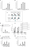
a, b Comparison of NTPase activities of free HelD and its complexes with RNAP. ATP/GTP hydrolyzing activity of free HelD was set as 100%. ATP hydrolysis (a) is stimulated upon complex formation, whereas GTP hydrolysis (b) remains almost unchanged. Control measurements for individual complex components are shown. The bars show averages from three biological replicates, the error bars are ±SD, the dots represent individual experiments (also in panels dg). c A scheme depicting the HelD release assay: His–RNAP was reconstituted into three different complexes, each containing combinations of HelD (cayn), σA (purple) and RbpA (yellow). The RNAP complexes were then allowed to bind to magnetic beads. The amount of HelD released, with or without addition of other factors (in panels dg) was determined by Coomassie blue-stained SDS-PAGE gels and densitometry. d Effect of 1 mM ATP, GTP, or CTP on HelD release. In panels dg, representative primary data are shown above the graph. Zero (Ø) shows HelD release without the addition of other factors. For this and experiments (e, g), the His–RNAP complex containing HelD, σA, and RbpA was used (depicted within the dashed box in c). The amount of HelD released from RNAP–σA–RbpA–HelD by the addition of ATP was set as 1 (also in other panels). A second primary data example is shown in Supplementary Fig. 14 together with a calibration curve used as quantification control. e, Effect of ATP analogs on HelD release. RNAP complexes were reconstituted as described in panel c with four HelD variants: WT-HelD (wild type), HelDσA-INT, HelDA-HYDRO, and HelDA-BIND (for definition of the mutants see Supplementary Fig. 16). Subsequently, 1 mM each of ATP, N-ATP, or ATPγS was added to the preformed RNAP complex attached to the magnetic beads and release of HelD from the complex was observed. f Release of HelD from the three types of complexes (c) induced with 1 mM ATP. g Effect of two forms of DNA and/or ATP on HelD release. CC, closed complex us-fork promoter DNA. OC open complex DNA with artificially opened transcription bubble. Source data are provided as a Source Data file.
Fig. 5
Fig. 5. Protective effect of HelD against rifampicin.
a Multiple round transcriptions from the Msm rRNA PrrnAPCL1 promoter were performed in the absence or presence of HelD with increasing amounts of rifampicin (RIF). The 1:1 RNAP:HelD ratio was used in protein reconstitution. Transcription at zero RIF was set as 1 for both ±HelD to facilitate visualization of the changes. The relative transcription in the absence of RIF and the presence of HelD compared to the absence of HelD was 72.4 % (lane 1). The bars show averages of three independent experiments, the dots are individual experimental data, the error bars show ±SD. p Values were calculated using a two-tailed, unpaired t-test. Source data are provided as the Source Data file. b HelD primary channel (PCh) loop binding causes conformational changes in the RNAP rifampicin binding site. Mtb RNAP–rifampicin complex (RIF in teal, RNAP β-core in light gray, PDB 5UHC) is superposed with the Msm us-fork–HelD–RPc-II complex (dark gray, active site Mg2+ in pink). Binding of the Msm HelD PCh loop (orange) deforms the RIF binding pocket: β-core/P483–N484 loop is pushed towards RIF by 2.7 Å, D485 of the HelD PCh loop itself sterically clashes with RIF, and β-core/R456, which usually coordinates RIF, is moved away. Possible atomic clashes between RIF and its deformed binding site and HelD are hinted with red ‘wave’ symbols, distances (green dashed lines) are in Å.
Fig. 6
Fig. 6. HelD participates during transcription initiation.
The circular arrangement model of HelD participation in transcription initiation is only the best approximation of the event succession displayed in panels ag: a HelD binds to the RNAP core together with σA and RbpA. Individual domains are color-coded according to the legend above. The HelD CO-domain interacts with σA2 and σAN-helix in the context of β′-clamp. In State I, the presence of full-length HelD in the primary channel (PCh) makes the β′-clamp wide open. However, it does not interfere with the AS and the RIF binding pocket. b In State II, the RNAP β′-clamp is maximally open and the HelD PCh-loop reaches the RNAP AS cavity and interferes with the RIF binding pocket. Concomitantly, in State II, the HelD–σA–RbpA–RNAP complex is able to recognize and bind DNA promoter outside the primary channel. c Disengagement of the HelD NTPase domain loosens the grip of the CO domain on the β′-clamp, the consequent narrowing of the primary channel tilts the CO domain. Still, the PCh-loop tip is folded into to AS cavity and interferes with the RIF binding pocket. d HelD clearance from the primary channel allows for β′-clamp closing, however HelDN-term presence in the secondary channel restricts the full closure. e Partial loading of dwDNA is compatible with HelD binding to the secondary channel, however, further interaction of dwDNA within the RNAP primary channel (f) triggers a conformational change of the secondary channel which disfavors and expels HelDN-term. σAN-helix helps to accommodate dwDNA towards the RP2-like intermediate. g In order to establish the RNAP open complex (competent for the first cycle of nucleotide addition), the DNA promoter still needs to be further accommodated into the RNAP AS cavity. At that moment, RNAP clamps around the dwDNA and σAN-helix locks the clamp by interaction with the β-lobe. From this state the complex can proceed towards transcription initiation or, when stalled by RIF, reverse towards HelD–RPc (gray dashed line arrow).

References

    1. Sutherland, C. & Murakami, K. S. An introduction to the structure and function of the catalytic core enzyme of Escherichia coli RNA polymerase. EcoSal Plus8, 1–9 (2018). - PMC - PubMed
    1. Paget, M. S. Bacterial sigma factors and anti-sigma factors: structure, function and distribution. Biomolecules5, 1245–1265 (2015). - PMC - PubMed
    1. Zhang, G. et al. Crystal structure of Thermus aquaticus core RNA polymerase at 3.3 A resolution. Cell98, 811–824 (1999). - PubMed
    1. Browning, D. F. & Busby, S. J. Local and global regulation of transcription initiation in bacteria. Nat. Rev. Microbiol.14, 638–650 (2016). - PubMed
    1. Wassarman, K. M. 6S RNA, a Global Regulator of Transcription. Microbiol. Spectr. 10.1128/microbiolspec.RWR-0019-2018 (2018) - PMC - PubMed

Publication types

MeSH terms

LinkOut - more resources