. 2025 Jan 3;16(1):362.
doi: 10.1038/s41467-024-55431-3.

Structural insights into the LGR4-RSPO2-ZNRF3 complexes regulating WNT/β-catenin signaling

Affiliations

Structural insights into the LGR4-RSPO2-ZNRF3 complexes regulating WNT/β-catenin signaling

Lu Wang et al. Nat Commun. .

Abstract

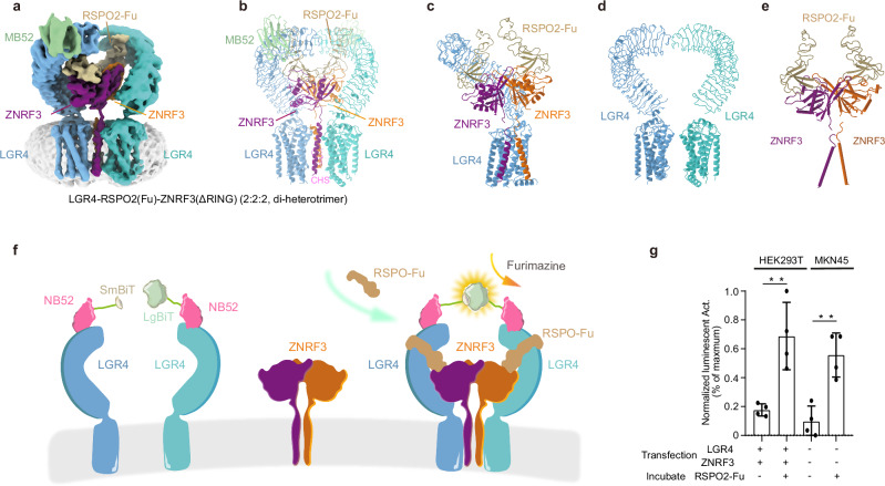
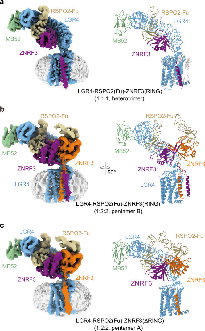
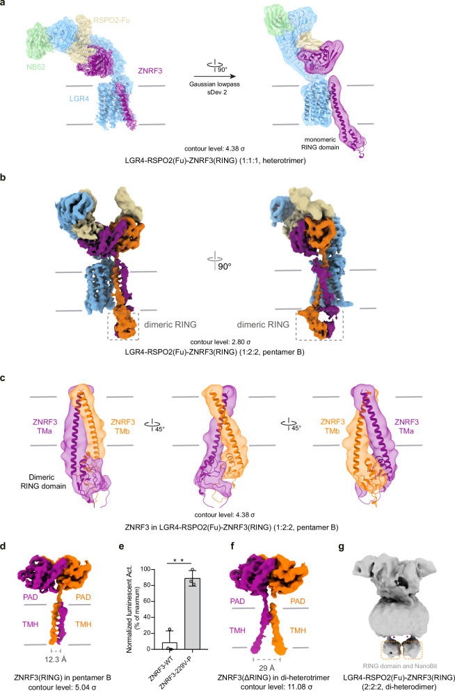
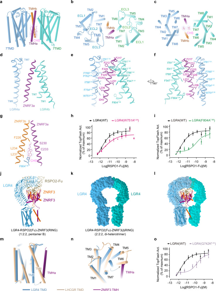
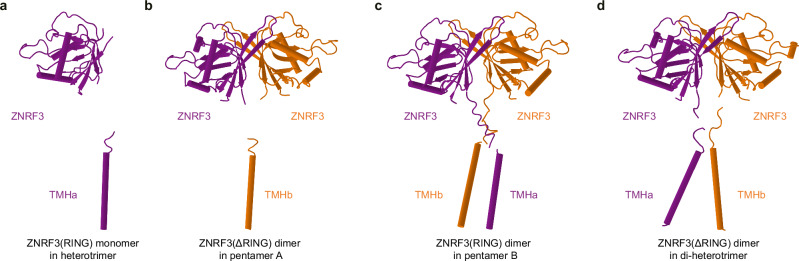
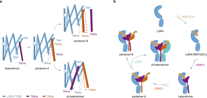
WNT/β-catenin signaling plays key roles in development and cancer1,2. ZNRF3/RNF43 modulates Frizzleds through ubiquitination, dampening WNT/β-catenin signaling. Conversely, RSPO1-4 binding to LGR4-6 and ZNRF3/RNF43 enhances WNT/β-catenin signaling3-5. Here, we elucidate the overall landscape of architectures in multiple LGR4, RSPO2, and ZNRF3 assemblies, showcasing varying stoichiometries and arrangements. These structures reveal that LGR4 and RSPO2 capture distinct states of ZNRF3. The intrinsic heterogeneity of the LGR4-RSPO2-ZNRF3 assembly is influenced by LGR4 content. Particularly, in the assembly complex with a 2:2:2 ratio, two LGR4 protomers induce and stabilize the inactive state of ZNRF3, characterized by a wide inward-open conformation of two transmembrane helices (TM helices). This specific assembly promotes a stable complex, facilitating LGR4-induced endocytosis of ZNRF3. In contrast, the active dimeric ZNRF3, bound by a single LGR4, adopts a coiled-coil TM helices conformation and dimerization of RING domains. Our findings unveil how LGR4 content mediates diverse assemblies, leading to conformational rearrangements in ZNRF3 to regulate WNT/β-catenin signaling, and provide a structural foundation for drug development targeting Wnt-driven cancers.


Conflict of interest statement

Competing interests: The authors declare no competing interests.

Figures

Fig. 1
Fig. 1. Cryo-EM structure of LGR4-RSPO2-ZNRF3(ΔRING) (2:2:2, di-heterotrimer) complex.
Structure of LGR4-RSPO2-ZNRF3(ΔRING) (2:2:2, di-heterotrimer) complex, Cryo-EM map (a) and atomic model (b) are shown. c The model of the di-heterotrimer complex (one LGR4 is omitted for clarity). d The arrangements of two LGR4 protomers in di-heterotrimer are shown from the front view. e The arrangements of RSPO2and ZNRF3 subunits are shown from the front view. Subunits LGR4, ZNRF3, and RSPO2 are colored in light blue/cyan, violet/orange, and brown, respectively. The nanobody portion of MB52 is shown in slate green (other segment is omitted for clarity). The same color scheme is used throughout the manuscript unless stated otherwise. f Schematic of the NanoBiT cell-based assay. The structure shows the proximity of two NB52s (pink). LgBiT (large subunit, light green) and SmBiT (small subunit, light brown) fragments are fused to the C-terminus of two NB52, respectively. The assembly of LGR4-RSPO2-ZNRF3(2:2:2) complex in the schematic (colored identically as in panel a) facilitates the luminescence complementation of NB52-LgBiT and NB52-SmBiT. g The results demonstrate a strong luminescent signal when RSPO2, NB52-LgBiT, and NB52-SmBiT, along with furimazine, are added to 293T cells (p = 0.005, n = 4) or MKN45 cancer cells (p = 0.0026, n = 4) expressing LGR4 and ZNRF3. Each value represents the mean ± SEM from four independent experiments. Source data are provided as a Source Data file.
Fig. 2
Fig. 2. Cryo-EM structures of LGR4-RSPO2ZNRF3(RING) (1:1:1, heterotrimer), LGR4-RSPO2-ZNRF3(RING) (1:2:2, pentamer B), LGR4-RSPO2-ZNRF3 (ΔRING) (1:2:2, pentamer A).
Structure of LGR4-RSPO2-ZNRF3(RING) (1:1:1, heterotrimer) (a), LGR4-RSPO2-ZNRF3(RING) (1:2:2, pentamer B) (b), and LGR4-RSPO2-ZNRF3(ΔRING) (1:2:2, pentamer A) (c), Cryo-EM map (left) and atomic model (right) are shown.
Fig. 3
Fig. 3. The rearrangement RING domain in different complexes.
a Superposition of the map and model of the LGR4-RSPO2-ZNRF3(RING) (1:1:1, heterotrimer, contour level: 2.8σ). b Map of the LGR4-RSPO2-ZNRF3(RING) (1:2:2, pentamer B, contour level: 2.8σ). c Superposition of the map and model of the TM helix and RING domain in pentamer B, shown in the low-pass map (contour level: 4.38σ) from different perspectives. d Map of ZNRF3(RING) in pentamer B (contour level: 5.4σ). e Wnt3a-stimulated-TOPFlash activity regulated by WT or V229-P-S230 mutant of ZNRF3, n = 4, p value = 0.0356. Source data are provided as a Source Data file. f Map of ZNRF3 (ΔRING) in di-heterotrimer (contour level: 11.08σ). g The map of the LGR4-RSPO2-ZNRF3 complex in its di-heterodimer form (2:2:2), with ZNRF3 containing the RING domain. Each value represents the mean ± SEM from four independent experiments.
Fig. 4
Fig. 4. LGR4 induces ZNRF3 into an inactive state.
The transmembrane interface between LGR4 and ZNRF3 in the LGR4-RSPO2-ZNRF3(ΔRING) complex (2:2:2, di-heterotrimer) is shown from the front view (a), top view (b) and bottom view (c). d The interface between the transmembrane domains of LGR4 and ZNRF3. e, f The side chain interactions between LGR4 and ZNRF3 within the transmembrane region of the LGR4-RSPO2(Fu)-ZNRF3(ΔRING) complex (2:2:2) are shown in detail. g The specific interactions between the transmembrane domain of LGR4, ZNRF3 and cholesteryl hemisuccinate within the LGR4-RSPO2(Fu)-ZNRF3(ΔRING) complex (2:2:2) are highlighted. h, i Dose-dependent TOPFlash activity induced by WT (black) or W751A6.39 mutant (red, e) and F804A7.56 mutant (green, f) of LGR4 after stimulation with RSPO1, n = 3. Source data are provided as a Source Data file. jl Comparing the LGR4 in pentamer B and di-heterotrimer. j The model of LGR4-RSPO2ZNRF3(RING) (1:2:2, pentamer B). k The surface of two LGR4 in LGR4-RSPO2ZNRF3(ΔRING) (2:2:2, di-heterotrimer). l Superposition of the LGR4 in pentamer B and di-heterotrimer (ZNRF3 and RSPO2 are deleted for clarity). m, n Conformational comparison of the TMD of LGR4 (light blue) in the di-heterotrimer with that of active LHCGR (wheat, PDB:7FII, RMSD = 1.524, 182 to 182 atoms) from the front view (m), and bottom view (n). The potential steric clash between TM6 of the active LGR4 and the single TM helix (violet) of ZNRF3 in the di-heterotrimer complex is shown. o TOPFlash plot illustrating the effect of breaking ionic lock (Q742K6.30 mutant) (purple) in the transmembrane domain of LGR4 on the activity of RSPO1, compared to that of WT (black), n = 4. Source data are provided as a Source Data file. Each value represents the mean ± SEM from independent experiments. The LGR4(WT) datasets in all three panels are identical.
Fig. 5
Fig. 5. Different conformational states of ZNRF3 in various assemblies of the LGR4-RSPO2(Fu)-ZNRF3 complexes.
a ZNRF3 in the heterotrimer: the linker between the extracellular domain of ZNRF3 and TM helix is absent. b ZNRF3 in pentamer A: one of the TM helices in the ZNRF3 dimer is completely missing, and the linker between the extracellular domain and the TM helix is also absent. c ZNRF3 in pentamer B: the two TM helices exhibit a “finger-crossed” arrangement. d ZNRF3 in the di-heterotrimer: the two TM helices adopt an inverted V-shaped configuration. ZNRF3 is depicted in purple or orange.
Fig. 6
Fig. 6. Proposed model for the LGR4-RSPO2-ZNRF3 complex assembly pathway.
a Front view of the transmembrane region of the LGR4-RSPO2-ZNRF3 in the different states aligned by the LGR4 TMD. The dashed line (violet/orange) indicates the position of ZNRF3 TM helices in the previous state. TM helix(a) is not visible in pentamer A, and the dashed line is colored in gray. For clarity, one LGR4 is omitted from the di-heterotrimer. b Cartoon illustrates the assembly pathway from the LGR4-apo state, and RSPO2-bound state, through LGR4-RSPO2-ZNRF3 (1:1:1, heterotrimer) state to LGR4-RSPO2-ZNRF3 (1:2:2, pentamer A) state, and then differentiates into the LGR4-RSPO2-ZNRF3 (1:2:2, pentamer B) state in the absence of LGR4, or into the LGR4-RSPO2-ZNRF3 (2:2:2, di-heterotrimer) state in the presence of LGR4.

References

    1. Nusse, R. & Clevers, H. Wnt/β-Catenin Signaling, Disease, and Emerging Therapeutic Modalities. Cell169, 985–999 (2017). - PubMed
    1. Steinhart, Z. & Angers, S. Wnt signaling in development and tissue homeostasis. Development145, 10.1242/dev.146589 (2018). - PubMed
    1. Hao, H. X. et al. ZNRF3 promotes Wnt receptor turnover in an R-spondin-sensitive manner. Nature485, 195–200 (2012). - PubMed
    1. Koo, B. K. et al. Tumour suppressor RNF43 is a stem-cell E3 ligase that induces endocytosis of Wnt receptors. Nature488, 665–669 (2012). - PubMed
    1. de Lau, W. et al. Lgr5 homologues associate with Wnt receptors and mediate R-spondin signalling. Nature476, 293–297 (2011). - PubMed

Publication types

MeSH terms

LinkOut - more resources