. 2024 Nov 3;15(1):9500.
doi: 10.1038/s41467-024-53923-w.

Structural diversity and clustering of bacterial flagellar outer domains

Affiliations

Structural diversity and clustering of bacterial flagellar outer domains

Jessie Lynda Fields et al. Nat Commun. .

Abstract

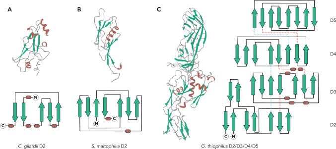
Supercoiled flagellar filaments function as mechanical propellers within the bacterial flagellum complex, playing a crucial role in motility. Flagellin, the building block of the filament, features a conserved inner D0/D1 core domain across different bacterial species. In contrast, approximately half of the flagellins possess additional, highly divergent outer domain(s), suggesting varied functional potential. In this study, we report atomic structures of flagellar filaments from three distinct bacterial species: Cupriavidus gilardii, Stenotrophomonas maltophilia, and Geovibrio thiophilus. Our findings reveal that the flagella from the facultative anaerobic G. thiophilus possesses a significantly more negatively charged surface, potentially enabling adhesion to positively charged minerals. Furthermore, we analyze all AlphaFold predicted structures for annotated bacterial flagellins, categorizing the flagellin outer domains into 682 structural clusters. This classification provides insights into the prevalence and experimental verification of these outer domains. Remarkably, two of the flagellar structures reported herein belong to a distinct cluster, indicating additional opportunities on the study of the functional diversity of flagellar outer domains. Our findings underscore the complexity of bacterial flagellins and open up possibilities for future studies into their varied roles beyond motility.


Conflict of interest statement

The authors declare no competing interests.

Figures

Fig. 1
Fig. 1. Cryo-EM of flagellar filaments from C. gilardii, S. maltophilia, and G. thiophilus.
Representative cryo-electron micrographs of the C. gilardii flagella (A), S. maltophilia flagella (B), and G. thiophilus flagella (C). Scale bar, 50 nm in (A)–(C). Orange arrowhead points to the C. gilardii flagellum; green arrowheads point to the S. maltophilia flagellum; blue arrowhead points to the G. thiophilus flagellum. 2D averages of all three flagella are shown in the top right corner. Cryo-EM reconstructions of the C. gilardii flagellum at 3.4 Å resolution (D), the S. maltophilia flagellum at 3.3 Å resolution (E), and the G. thiophilus flagellum at 4.1 Å resolution (F). Thin sections parallel to the helical axis of the flagella are shown on the right of the 3D reconstruction, colored by the radius. G–I The single flagellin structures modeled into the cryo-EM densities are displayed on the right, with their respective domain names indicated. The conserved D0/D1 domains are shown in gray. The outer domains of C. gilardii, S. maltophilia, and G. thiophilus are consistently colored in accordance with the 3D reconstructions.
Fig. 2
Fig. 2. The fold of flagellar outer domains from C. gilardii, S. maltophilia, and G. thiophilus.
Cryo-EM structures of the flagellar outer domains from C. gilardii (A), S. maltophilia (B), and G. thiophilus (C) were presented, with all α-helices colored brown and β-sheets colored green. These domains are also drawn in schematic representation, with β-sheets shown as green arrows, α-helices as brown cylinders, and loops as dark gray lines. When multiple domains are present, they are connected by blue dashed lines near the N-terminus and red dashed lines near the C-terminus.
Fig. 3
Fig. 3. Outer domain arrangements on flagellar surface.
A The top view of three flagellar filaments with the conserved D0/D1 domains in gray, the D2 domain of C. gilardii flagellum in orange, the D2 domain of S. maltophilia flagellum in green, the D2/D3 domains of G. thiophilus flagellum in light blue, and the D4/D5 domains of G. thiophilus flagellum in dark blue. B The D2 domain dimerization in C. gilardii is presented within the cryo-EM map density. C The D4/D5 domain dimerization in G. thiophilus, with the model shown within cryo-EM map density. D The helical net of the flagellar filament is illustrated using the convention that the surface is unrolled, providing a view from the outside. Gray dots indicate the approximate symmetry present in the D0/D1 domains of all flagellar filaments. The conventional 11-start and left-handed 5-start, originating from the D0/D1 domain, are indicated with black lines. The dimer interface that occurs in the flagellar outer domains of both C. gilardii and G. thiophilus is illustrated with transparent orange stadium shapes. The seam arising from this dimerization packed along the D0/D1 5-start is indicated by a red dashed line.
Fig. 4
Fig. 4. Analysis of AlphaFold predictions of bacterial flagellin outer domains.
A The typical architecture of flagellin includes outer domains. The D0 and D1 domains, which have 263 or a bit more residues, are colored in red and purple, respectively. The other outer domains, extending from inner to outer diameter, are sequentially named D2, D3, D4, and so on. B This plot represents the number of UniProt entries corresponding to various lengths of flagellin proteins. Flagellin AlphaFold predictions equal and longer than 350 residues are selected for subsequent multistep analysis, as described in the method section. 682 structural clusters were generated. C The matrix visualizes a DALI all-to-all analysis of 682 structural cluster representatives. It is constructed using pairwise DALI Z-scores, with the corresponding Z-score color scale presented on the right. The most prominent cluster, located at the bottom right of the matrix, contains representatives with Ig-like domains.
Fig. 5
Fig. 5. Populated flagellin outer domains in the bacterial domain.
A The relationships among 682 structural clusters are depicted using the Fruchterman-Reingold algorithm, a force-directed layout algorithm from the R/igraph package. The communities, indicated with transparent color circles, were detected using the walktrap algorithm with a step size of 6. Additionally, some most populated clusters, as illustrated in (B), are highlighted with black circles. B AlphaFold predictions or experimentally determined structures from populous structural clusters are organized according to the population size of the first representative in a given community. Within these models, all α-helices are colored yellow, β-sheets blue, and loops gray. Known domain folds are labeled. For clusters with available cryo-EM or X-ray structures, the corresponding species and PDB IDs are provided.

Update of

References

    1. Soutourina, O. A., Semenova, E. A., Parfenova, V. V., Danchin, A. & Bertin, P. Control of bacterial motility by environmental factors in polarly flagellated and peritrichous bacteria isolated from Lake Baikal. Appl. Environ. Microbiol.67, 3852–3859 (2001). - PMC - PubMed
    1. Zhou, B. B., Szymanski, C. M. & Baylink, A. Bacterial chemotaxis in human diseases. Trends Microbiol.31, 453–467 (2023). - PMC - PubMed
    1. Colin, R., Ni, B., Laganenka, L. & Sourjik, V. Multiple functions of flagellar motility and chemotaxis in bacterial physiology. FEMS Microbiol. Rev.45, 10.1093/femsre/fuab038 (2021). - PMC - PubMed
    1. Spohn, G., and Scarlato, V. Motility, Chemotaxis, and Flagella. In Helicobacter pylori: Physiology and Genetics (eds Mobley, H. L. T., Mendz, G. L. & Hazell, S. L.) (2001). - PubMed
    1. Palma, V., Gutierrez, M. S., Vargas, O., Parthasarathy, R. & Navarrete, P. Methods to evaluate bacterial motility and its role in bacterial–host interactions. Microorganisms10, 10.3390/microorganisms10030563 (2022). - PMC - PubMed

Publication types