. 2024 Sep 7;15(1):7839.
doi: 10.1038/s41467-024-52130-x.

Burkholderia cenocepacia epigenetic regulator M.BceJIV simultaneously engages two DNA recognition sequences for methylation

Affiliations

Burkholderia cenocepacia epigenetic regulator M.BceJIV simultaneously engages two DNA recognition sequences for methylation

Richard Quintana-Feliciano et al. Nat Commun. .

Abstract

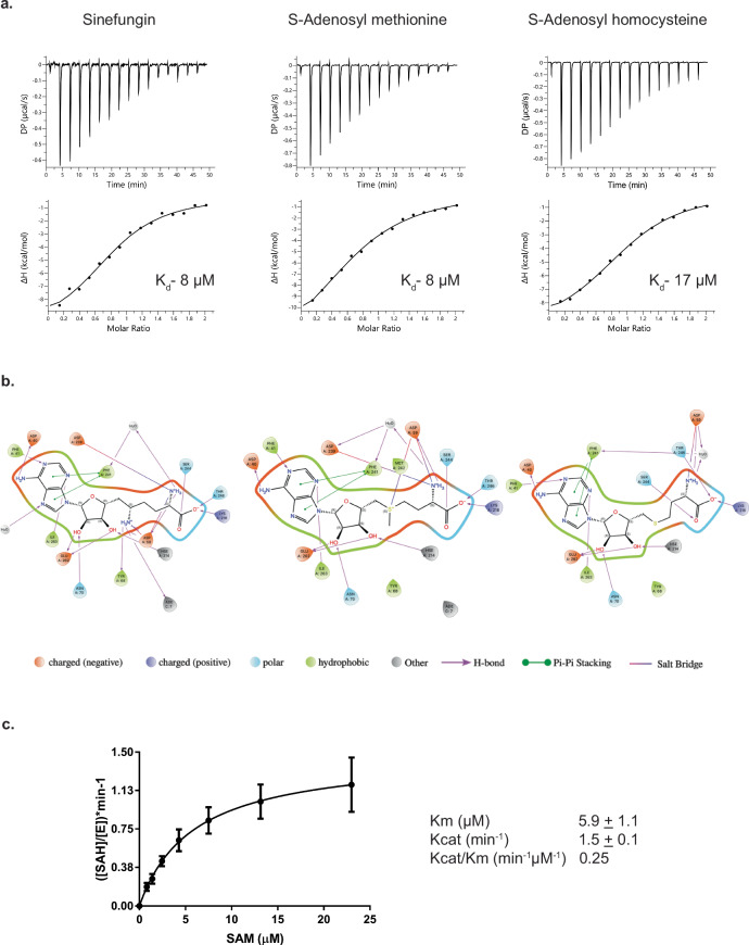
Burkholderia cenocepacia is an opportunistic and infective bacterium containing an orphan DNA methyltransferase called M.BceJIV with roles in regulating gene expression and motility of the bacterium. M.BceJIV recognizes a GTWWAC motif (where W can be an adenine or a thymine) and methylates N6 of the adenine at the fifth base position. Here, we present crystal structures of M.BceJIV/DNA/sinefungin ternary complex and allied biochemical, computational, and thermodynamic analyses. Remarkably, the structures show not one, but two DNA substrates bound to the M.BceJIV dimer, with each monomer contributing to the recognition of two recognition sequences. We also show that methylation at the two recognition sequences occurs independently, and that the GTWWAC motifs are enriched in intergenic regions in the genomes of B. cenocepacia strains. We further computationally assess the interactions underlying the affinities of different ligands (SAM, SAH, and sinefungin) for M.BceJIV, as a step towards developing selective inhibitors for limiting B. cenocepacia infection.


Conflict of interest statement

The authors declare no competing interests.

Figures

Fig. 1
Fig. 1. Overall structure of M.BceJIVΔ29-DNA-sinefungin complex.
M.BceJIV forms a homodimer complex with two DNAs and two sinefungin molecules. Monomer A (magenta) receives the adenine to be methylated from DNA1, but its TRD reaches over to DNA2 and, conversely, monomer B (cyan) receives the adenine to be methylated from DNA2, but its TRD extends over to DNA1. Helices are labeled from αA to αH, and strands are labeled from β1–β8. Sinefungin is shown in a stick representation. The DNA substrate used for crystallization is shown, and the recognition sequence is boxed and numbered.
Fig. 2
Fig. 2. Structure of M.BceJIVΔ29 monomers bound to DNA and sinefungin, and the electron density for DNA and sinefungin molecules.
Structure of monomer A bound to DNA1 and sinefunginA (a) and of monomer B bound to DNA2 and sinefunginB (b) showing DNA distortions and the TRDs. Each monomer contributes to the recognition of its own methylation-target DNA via its loop-45 and loop-6F through the minor groove and to the recognition of a separate DNA via its TRD through the major groove. 2Fo-Fc map contoured at 1.5 σ is shown for DNA1 and sinefunginA (c) and for DNA2 and sinefunginB (d). The recognition sequence is labeled.
Fig. 3
Fig. 3. Molecular basis of target (DNA1) recognition.
a The first base pair (G1:C1) is specified by contacts with Gln207 of loop-6F of monomer A and Arg180 of TRDB; the second base pair (T2:A2) makes contact with His205 of loop-6F of monomer A and van der Waals contacts with Arg180 of TRDB; the third base pair (T3:A3) is specified by contacts with His205 and Arg203 of loop-6F of monomer A and van der Waals contacts with Leu182 and Phe183 of TRDB; the fourth base pair (A4:T4) makes contacts with Arg203 and van der Waals contacts with Met136 of monomer A (from loop-45) and van der Waals contacts with Trp188 of TRDB; at the fifth base pair (A5:T5), the thymine opposite the adenine to be methylated is specified by contacts with Gly137 of loop-45 that embeds in place of the everted fifth adenine and sixth cytosines of the target strand, and van der Waals contacts with Trp188 of TRDB; at the sixth base pair (C6:G6), the guanine opposite to the everted cytosine makes contacts with Gly137 of loop-45 of monomer A. b A close-up view of the flipped-out adenine that is accommodated in the catalytic cleft of monomer A and makes contacts with Asp59, Pro60 and Tyr62 of the conserved DPPY motif and with Tyr68 and Lys218 of monomer A. c A close-up view of the flipped-out cytosine accommodated outside the catalytic cleft and makes contacts with Asp148, His147, Thr106, Trp107, and Gln108 of monomer A. Monomer A is colored as magenta and the TRDB is colored as cyan.
Fig. 4
Fig. 4. Interactions of M.BceJIVΔ29 with sinefungin and the flipped adenine.
A close-up view of specific M.BceJIVΔ29-sinefungin interactions and residues shared between sinefungin and the flipped-out adenine. Sinefungin is tightly bound within the MTase domain via extensive hydrogen bonds (dashed lines) and hydrophobic contacts.
Fig. 5
Fig. 5. M.BceJIV binds two DNA sites in solution and methylates them independently.
a Results from sedimentation velocity analysis showing the molecular weight distribution of M.BceJIVΔ29 in the absence of DNA (blue) and in the presence of DNA (red). The small peaks at the low end of the distribution are small contaminants, as confirmed by SDS-PAGE. The experiments were repeated in duplicates or triplicates, and the average molecular weight was reported. b Addition of a second DNA methylation site in trans (left) or in cis (right) does not assist the methylation of the first DNA site. In the trans experiment (left), HpaI digestion of 24 nM BamHI-linearized pUC18 containing a single HpaI site with increasing concentrations (0 nM to 50 nM) of a 19 mer DNA duplex in trans containing an M.BceJIV methylation motif and no HpaI site in the presence of 24 nM M.BceJIV and 20 μM SAM in a 50 s reaction. In the cis experiment (right), HpaI digestion of 24 nM BamHI-linearized pUC18 containing a single HpaI site (or both an HpaI site and another M.BceJIV methylation site in cis) with increasing reaction time (1 min to 5 min) in the presence of 24 nM or 48 nM M.BceJIV and 20 μM SAM. The experiments were repeated twice with similar results.
Fig. 6
Fig. 6. Distribution of GTWWAC motif sites across two strains of B. cenocepacia.
a, b Circos plot showing the genome-wide distribution of GTWWAC sites along the three chromosomes and a plasmid of B. cenocepacia J2315 and B. cenocepacia K56-2, respectively. c, d Density plot for the distribution of distances between each two neighboring GTWWAC sites in B. cenocepacia strains J2315 and K56-2. e, f Enrichment of GTWWAC in the intergenic regions (observed frequency vs. expected frequency) in the two strains.
Fig. 7
Fig. 7. ITC analysis of M.BceJIVΔ29 with SAM, SAH and sinefungin, and kinetics of M.BceJIV using a bioluminescence assay.
a ITC titration data for sinefungin (left), SAM (middle), and SAH (right) with M.BceJIVΔ29. The equilibrium dissociation constants (KD) were derived from the resulting binding isotherms. All the experiments were repeated in duplicates and the average value is reported. b Two-dimensional protein-ligand interaction diagrams generated using energy-minimized conformations of the crystal structure of M.BceJIVΔ29 bound to sinefungin (left) and predicted structures of SAM and SAH in M.BceJIVΔ29 (middle and right, respectively) embedded in an explicit water environment. The Ligand Interaction script in Maestro (Schrödinger Inc., http://www.schrodinger.com/) was used to generate these diagrams with default parameters. Among the protein residues within a 4 Å distance of the ligands, only those that interact with the ligands according to Maestro’s default parameters (see details in Supplementary Table 2) are displayed. c Kinetics of SAH byproduct formation by varying concentrations of the methyl donor SAM. A summary of kinetic parameters is also shown. Data are presented as mean values +/− SD from experiments performed in triplicates, and the source data are provided as a Source Data file.

Update of

References

    1. Loutet, S. A. & Valvano, M. A. A decade of Burkholderia cenocepacia virulence determinant research. Infect. Immun.78, 4088–4100 (2010). 10.1128/IAI.00212-10 - DOI - PMC - PubMed
    1. Roberts, R. J., Vincze, T., Posfai, J. & Macelis, D. REBASE–enzymes and genes for DNA restriction and modification. Nucleic Acids Res.35, D269–D270 (2007). 10.1093/nar/gkl891 - DOI - PMC - PubMed
    1. Beaulaurier, J., Schadt, E. E. & Fang, G. Deciphering bacterial epigenomes using modern sequencing technologies. Nat. Rev. Genet.20, 157–172 (2019). 10.1038/s41576-018-0081-3 - DOI - PMC - PubMed
    1. Oliveira, P. H. & Fang, G. Conserved DNA methyltransferases: A window into fundamental mechanisms of epigenetic regulation in bacteria. Trends Microbiol.29, 28–40 (2021). 10.1016/j.tim.2020.04.007 - DOI - PMC - PubMed
    1. Zhou, J., Horton, J. R., Blumenthal, R. M., Zhang, X. & Cheng, X. Clostridioides difficile specific DNA adenine methyltransferase CamA squeezes and flips adenine out of DNA helix. Nat. Commun.12, 3436 (2021). 10.1038/s41467-021-23693-w - DOI - PMC - PubMed

Publication types

MeSH terms

LinkOut - more resources