. 2005 Oct 1;61(Pt 10):867-74.
doi: 10.1107/S1744309105029842. Epub 2005 Sep 30.

Structures of a putative RNA 5-methyluridine methyltransferase, Thermus thermophilus TTHA1280, and its complex with S-adenosyl-L-homocysteine

Affiliations

Structures of a putative RNA 5-methyluridine methyltransferase, Thermus thermophilus TTHA1280, and its complex with S-adenosyl-L-homocysteine

Augen A Pioszak et al. Acta Crystallogr Sect F Struct Biol Cryst Commun. .

Abstract

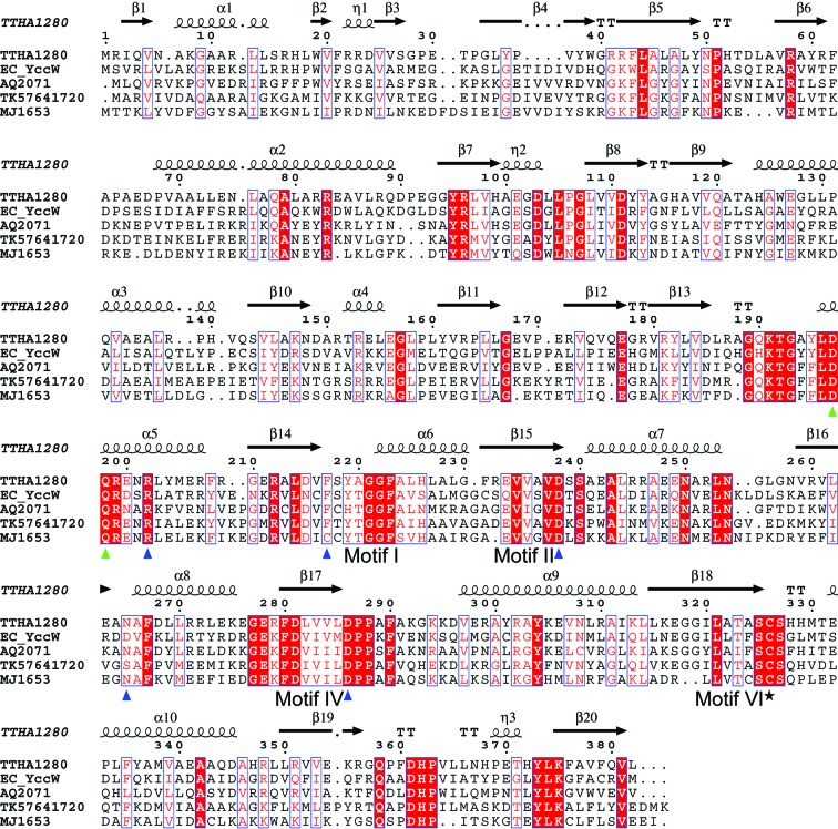
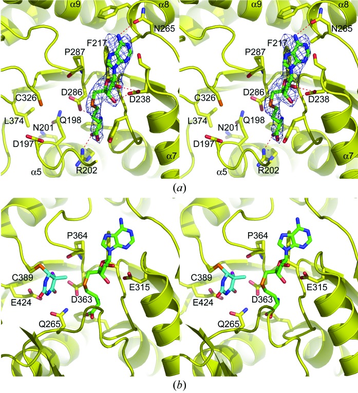
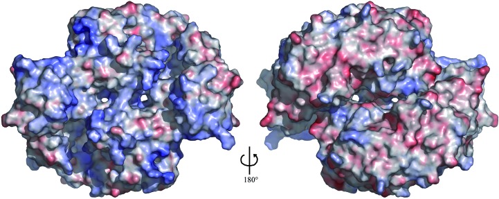
The Thermus thermophilus hypothetical protein TTHA1280 belongs to a family of predicted S-adenosyl-L-methionine (AdoMet) dependent RNA methyltransferases (MTases) present in many bacterial and archaeal species. Inspection of amino-acid sequence motifs common to class I Rossmann-fold-like MTases suggested a specific role as an RNA 5-methyluridine MTase. Selenomethionine (SeMet) labelled and native versions of the protein were expressed, purified and crystallized. Two crystal forms of the SeMet-labelled apoprotein were obtained: SeMet-ApoI and SeMet-ApoII. Cocrystallization of the native protein with S-adenosyl-L-homocysteine (AdoHcy) yielded a third crystal form, Native-AdoHcy. The SeMet-ApoI structure was solved by the multiple anomalous dispersion method and refined at 2.55 A resolution. The SeMet-ApoII and Native-AdoHcy structures were solved by molecular replacement and refined at 1.80 and 2.60 A, respectively. TTHA1280 formed a homodimer in the crystals and in solution. Each subunit folds into a three-domain structure composed of a small N-terminal PUA domain, a central alpha/beta-domain and a C-terminal Rossmann-fold-like MTase domain. The three domains form an overall clamp-like shape, with the putative active site facing a deep cleft. The architecture of the active site is consistent with specific recognition of uridine and catalysis of methyl transfer to the 5-carbon position. The cleft is suitable in size and charge distribution for binding single-stranded RNA.


Figures

Figure 1
Figure 1
Amino-acid sequence alignment of selected MJ1653 family proteins from bacterial and archaeal species. The secondary-structure elements of TTHA1280 are shown above the sequence. Identical residues are highlighted with a red background and similar residues are shown in red text with a white background. The putative catalytic cysteine residue is indicated with a black star, residues that form hydrogen bonds or hydrophobic contacts with the AdoHcy ligand are indicated with blue triangles and residues with possible roles in substrate base recognition (as described in text) are indicated with green triangles. The alignment was performed with CLUSTALW (Thompson et al., 1994 ▶) and the figure was generated with ESPript (Gouet et al., 1999 ▶). The bacterial species are Thermus thermophilus (TTHA), Escherichia coli (EC) and Aquifex aeolicus (AQ). The archaeal species are Thermococcus kodakaraensis (TK) and Methanococcus jannaschii (MJ).
Figure 2
Figure 2
Catalytic mechanism of AdoMet-dependent RNA m5U MTases (adapted from Kealey et al., 1994 ▶).
Figure 3
Figure 3
Overall structure of the TTHA1280 apoprotein. (a) Ribbon diagram of a single subunit of the apoprotein presented in stereo with secondary-structure elements labelled. The N-terminal, central and C-terminal domains are colored cyan, lime and yellow, respectively. The putative catalytic Cys326 is shown in stick representation and coloured red. (b) Ribbon diagram of the apoprotein homodimer viewed down the non-crystallographic twofold axis. (c) Molecular surface of a single subunit of the apoprotein coloured by amino-acid sequence conservation. A semi-transparent surface is superimposed on a ribbon diagram oriented as in (a). The strictly conserved residues shown in Fig. 1 ▶ are coloured cyan, the similar residues are coloured green and the unconserved residues are coloured white. The square highlights the putative active site. This and all subsequent figures were generated with PyMOL (DeLano, 2002 ▶).
Figure 4
Figure 4
Comparison of TTHA1280 and E. coli RumA. A single TTHA1280 subunit is shown on the left, next to the RumA monomer (PDB code 1uwv). The proteins are positioned with their C-terminal domains (yellow) in the same orientation. The RumA N-terminal domain is coloured red and secondary-structure elements present in RumA, but not in TTHA1280, are coloured grey. The portion of the RumA central domain that is equivalent to the TTHA1280 central domain is colored lime for elements that exhibit the same topology as in TTHA1280 and pink for elements with topology different to that in TTHA1280. The RumA 4Fe–4S cluster is shown in stick representation. Selected secondary-structure elements are labelled as in Fig. 3 ▶(a) for TTHA1280 or as in Lee et al. (2004 ▶) for RumA. The squares highlight the putative (TTHA1280) and experimentally confirmed (RumA) active sites.
Figure 5
Figure 5
AdoHcy binding and comparison of the TTHA1280 and RumA active sites. (a) Stereoview of the putative TTHA1280 active site from the 2.6 Å resolution Native-AdoHcy structure. The protein is shown as a ribbon diagram with selected side chains and the AdoHcy ligand displayed as sticks. Atoms are coloured red for oxygen, blue for nitrogen, orange for sulfur, yellow for protein C atoms and green for AdoHcy C atoms. The red dashes indicate hydrogen bonds between the protein and AdoHcy. The simulated-annealing composite omit 2F oF c electron-density map is shown as a blue mesh contoured at 1σ and displayed within 2 Å of the ligand. (b) Stereoview of the RumA active site from the RumA–RNA–AdoHcy ternary complex (PDB code 2bh2). For clarity, only the uracil base of the RNA substrate is shown. The base is methylated at the C5 position and has an F atom in place of the C5 hydrogen, preventing β-elimination of the enzyme (Lee et al., 2005 ▶). Uracil C atoms are coloured cyan and the F atom is coloured magenta.
Figure 6
Figure 6
Electrostatic surface potential of the TTHA1280 apoprotein homodimer. The left image shows the same view as in Fig. 3 ▶(b). Positively charged regions are coloured blue, negatively charged regions red and neutral regions white. The colour ramp is from −8 to +8kT, where k is the Boltzmann constant and T is the absolute temperature. The surface potential was calculated with APBS (Baker et al., 2001 ▶) assuming a solvent of 150 mM NaCl. The molecular surface was generated with PyMOL (DeLano, 2002 ▶).

References

    1. Agarwalla, S., Kealey, J. T., Santi, D. V. & Stroud, R. M. (2002). J. Biol. Chem. 277, 8835–8840. - PubMed
    1. Anantharaman, V., Koonin, E. V. & Aravind, L. (2002). Nucleic Acids Res. 30, 1427–1464. - PMC - PubMed
    1. Aravind, L. & Koonin, E. V. (1999). J. Mol. Evol. 48, 291–302. - PubMed
    1. Baker, N. A., Sept, D., Joseph, S., Holst, M. J. & McCammon, J. A. (2001). Proc. Natl Acad. Sci. USA, 98, 10037–10041. - PMC - PubMed
    1. Brünger, A. T., Adams, P. D., Clore, G. M., DeLano, W. L., Gros, P., Grosse-Kunstleve, R. W., Jiang, J.-S., Kuszewski, J., Nilges, M., Pannu, N. S., Read, R. J., Rice, L. M., Simonson, T. & Warren, G. L. (1998). Acta Cryst. D54, 905–921. - PubMed

Publication types

MeSH terms